DHEA-S blood test: normal by age, high and low causes
DHEA-S is an adrenal hormone that falls with age. Here's the normal range by age and sex, what high and low levels usually mean, and when it's urgent.
DHEA-S (dehydroepiandrosterone sulfate) is a hormone your adrenal glands make in large amounts, and your body turns it into testosterone and estrogen where it needs them. It peaks in your 20s and then falls steadily, so a normal DHEA-S depends on your age and sex.12 A high level most often goes with polycystic ovary syndrome (PCOS) in women.3 A low level usually reflects age; steroid medicines and, rarely, adrenal gland failure are the other common causes.45
The short version
- Normal depends on age and sex. MedlinePlus lists typical ranges of 110–510 µg/dL for men and 45–320 µg/dL for women aged 18–29, falling to under 204 (men) and under 145 (women) after 59.4 Use the range on your own report.
- Mildly high, most often in a young woman with acne, extra hair or irregular periods, usually points to PCOS. About 20–30% of women with PCOS have a high DHEA-S.36
- Over 700 µg/dL in a woman with signs of excess male hormones is the level at which endocrinologists rule out an adrenal tumor. That needs prompt medical care.7
- Low is expected with age and common with steroid medicines.14 A DHEA-S well below the range for your age can also be an early clue to adrenal insufficiency, which is confirmed with cortisol and ACTH tests.5
What DHEA-S is
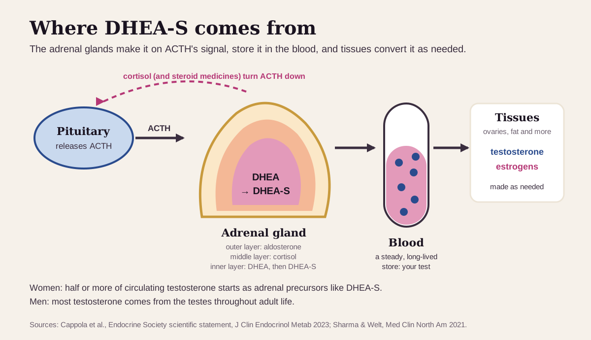
Your adrenal glands sit on top of your kidneys. Their inner layer, the zona reticularis, makes DHEA and then attaches a sulfate group to it, which turns it into DHEA-S. The same pituitary signal that drives cortisol, ACTH, drives DHEA-S too.1 Smaller amounts come from the testicles and ovaries.2
DHEA-S is mostly a storage form. On its own it acts only weakly on androgen receptors. The ovaries, fat and other tissues convert it into testosterone and estrogens as they need them.793 (More on the chemistry at the end.)
That makes DHEA-S more important for women. Half or more of a woman's circulating testosterone starts as adrenal precursors such as DHEA-S, while most of a man's comes from the testes.1 DHEA-S is also the best single marker of excess adrenal androgens in most situations.7
Doctors usually order it to look into extra facial or body hair, acne, irregular periods or a deepening voice in women, early or late puberty in boys, or a suspected adrenal problem. It's often run alongside testosterone and estrogen tests.2
What's a normal DHEA-S?
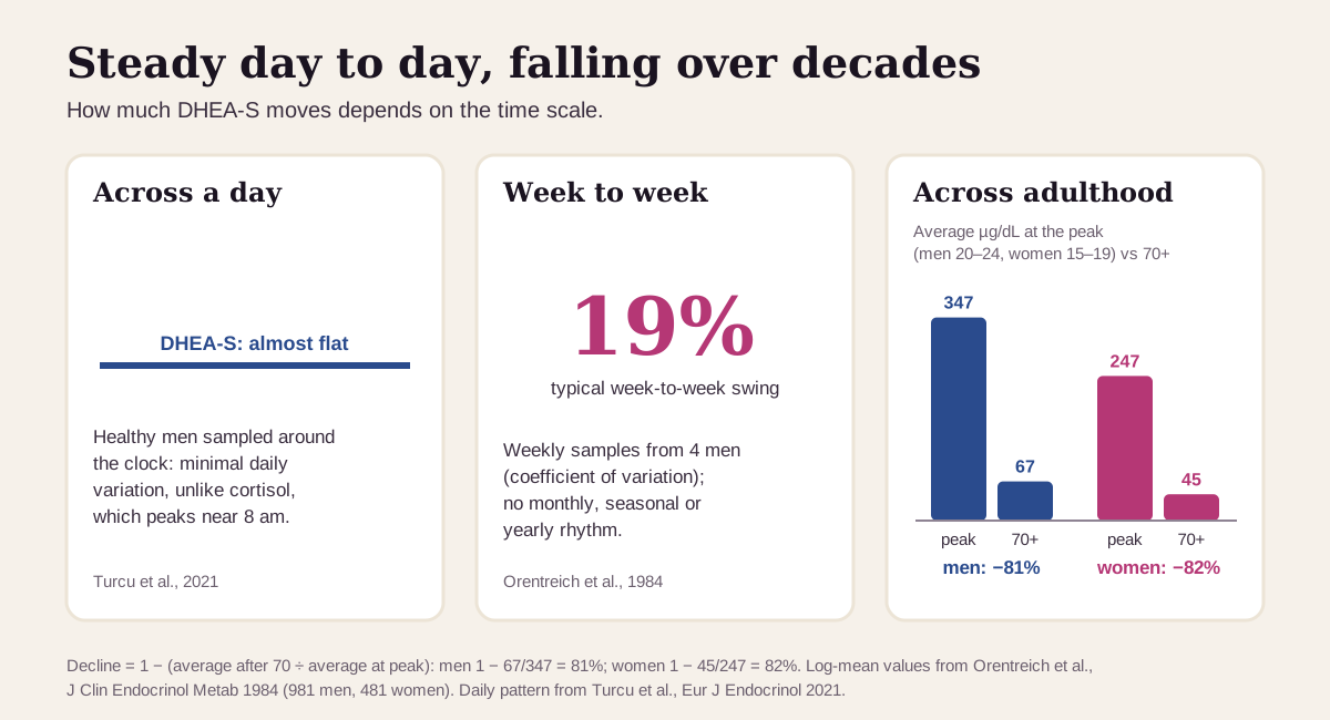
Your age and sex set the range. DHEA-S rises from about age 7–8 (a stage called adrenarche), peaks in your 20s, and falls to childhood levels by about age 80 in most adults.71 In a study of 981 men and 481 women, average DHEA-S peaked at 347 µg/dL in men aged 20–24 and 247 µg/dL in women aged 15–19. After 70 it averaged 67 and 45 µg/dL. Men ran higher than women at every age from 20 to 69.10
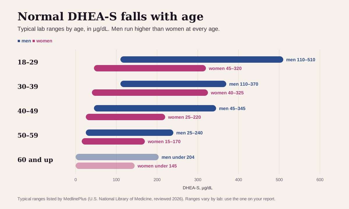
- Ages 18–29: 110–510 µg/dL for men, 45–320 µg/dL for women.
- Ages 40–49: 45–345 µg/dL for men, 25–220 µg/dL for women.
- Over 59: under 204 µg/dL for men, under 145 µg/dL for women.4
Ranges vary slightly between labs, which use different measurements.4 The range printed on your report is the one to use, and comparing results works best from the same lab.
"Optimal" DHEA-S: what health optimizers aim for
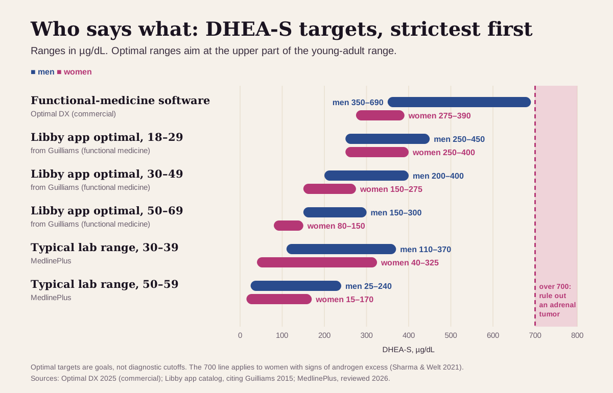
Functional-medicine references aim for the upper part of the young-adult range, well above what a lab calls normal for your age.1112 Here's the ladder, from strictest to loosest:
| Target | Men | Women | Who uses it |
|---|---|---|---|
| Strictest (functional-medicine software) | 350–690 µg/dL | 275–390 µg/dL | Optimal DX's published optimal range, with no age adjustment11 |
| Optimal range in the Libby app, ages 18–29 | 250–450 µg/dL | 250–400 µg/dL | Age- and sex-specific functional ranges, from a functional-medicine textbook on the adrenal (HPA) axis12 |
| Libby app, ages 30–49 | 200–400 µg/dL | 150–275 µg/dL | Same source12 |
| Libby app, ages 50–69 | 150–300 µg/dL | 80–150 µg/dL | Same source12 |
| Endocrine Society treatment target | — | mid-range for premenopausal women | For women with adrenal insufficiency who take DHEA5 |
| Typical lab range, ages 30–39 | 110–370 µg/dL | 40–325 µg/dL | MedlinePlus4 |
| Urgent | — | over 700 µg/dL | Rule out an adrenal tumor promptly7 |
Why optimizers watch a low DHEA-S:
- In 2,644 Swedish men aged 69–81 followed for 4.5 years, those in the lowest quarter of DHEA-S had a 54% higher risk of death from any cause than the other men (hazard ratio 1.54), and a 61% higher risk of cardiovascular death, after adjusting for the usual heart risk factors.13
- The link was strongest in the younger half of that group: an age-adjusted 2.64 times higher risk of cardiovascular death in men under 75.4.13
- In women, higher DHEA-S also appears to go with lower cardiovascular risk.6
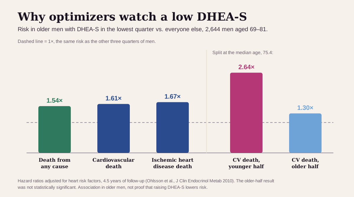
Keep "optimal" in perspective. A low DHEA-S tracks with age and illness, and raising the number with pills hasn't delivered the hoped-for benefits. In a 2-year randomized trial, DHEA raised DHEA-S by a median of about 340 µg/dL in older men and 380 µg/dL in older women. Body composition, strength, insulin sensitivity and quality of life stayed the same; the only gains were in bone density at single sites.14 The Endocrine Society recommends against routine DHEA use in women, and its statement on aging concludes the trials give no support for DHEA as an anti-aging supplement.151 Higher is also not always better: a high DHEA-S goes with PCOS and, rarely, a tumor.3
What a high DHEA-S usually means
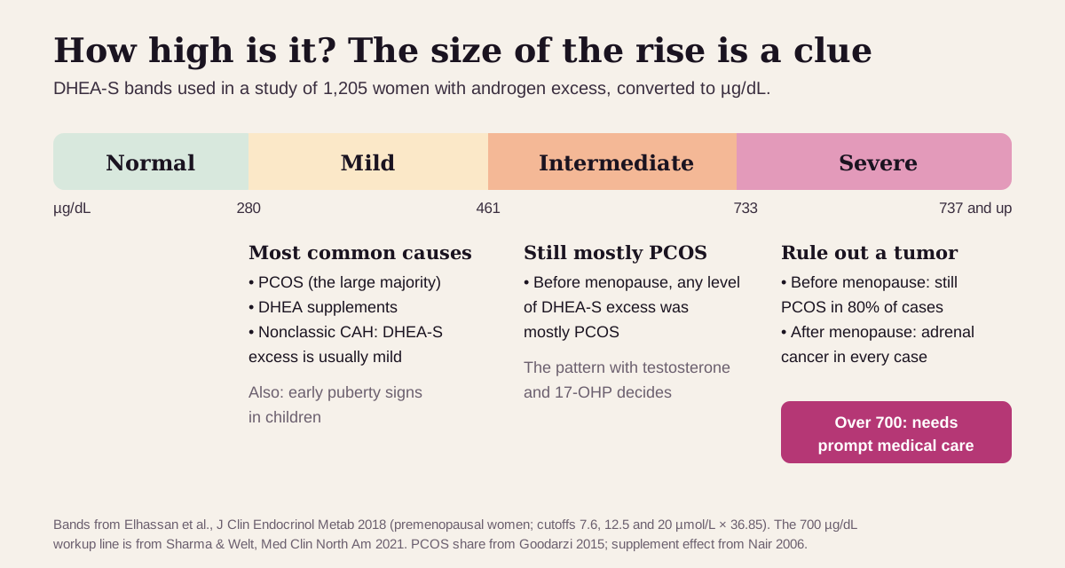
The size of the rise is the first clue. In a study of 1,205 women with androgen excess, researchers graded DHEA-S into mild, intermediate and severe bands.3
Mildly high: common, usually slow-moving
The usual suspects:
- PCOS. It's the most common cause of excess male hormones in women: 89% of premenopausal women with a high androgen in the 1,205-woman study had PCOS.3 About 20–30% of women with PCOS have a high DHEA-S.6
- DHEA supplements. Over-the-counter DHEA raises DHEA-S a lot, by a median of 340–380 µg/dL in one trial.14 It's sold over the counter as a dietary supplement, outside FDA regulation.1 Tell your clinician if you take it.
- Nonclassic congenital adrenal hyperplasia (CAH). An inherited shortfall in one adrenal enzyme (21-hydroxylase), found in about 1 in 200 people in the U.S.16 It often gets mistaken for PCOS. DHEA-S may be normal or only mildly high (in one clinic series, DHEA-S excess in CAH was less common and always mild), and an early-morning 17-hydroxyprogesterone (17-OHP) test is the clue.37
- Less common: early puberty changes in children.4
In men, a high DHEA-S may cause no symptoms at all.2
Very high (over 700 µg/dL): rule out a tumor
Adrenal tumors cause no more than 0.2% of cases of androgen excess, but they matter. DHEA-S was high in 82% of women with androgen-secreting adrenal tumors.17 Endocrinologists rule out an adrenal tumor when DHEA-S is over 700 µg/dL, usually with an adrenal CT scan.7 Before menopause, even severe DHEA-S excess was still PCOS 80% of the time. After menopause, every case of severe DHEA-S excess in the Birmingham study was adrenal cancer.3
Get prompt care if a high DHEA-S comes with fast-growing facial or body hair, a deepening voice, balding at the crown or a sudden gain in muscle, especially when the changes come on over months, or if your DHEA-S is high after menopause.23
What a low DHEA-S usually means
- Age. The most common reason. After 70, average DHEA-S is about a fifth of its peak.10 Older adults can have a low DHEA-S with healthy adrenal glands.5
- Steroid medicines. Prednisone-type pills lower DHEA-S.4 Even medium-to-high doses of inhaled steroids can: in 22 children with asthma on them, 13 had a weak cortisol response, and their DHEA-S ran lower, which makes it a useful screen.18 Keep taking a prescribed steroid and bring the result to your clinician.
- Adrenal insufficiency or a pituitary problem. The Endocrine Society calls a DHEA-S well below the lower limit for your age and sex a useful early sign of adrenal insufficiency (Addison's disease). The diagnosis is then made with a morning cortisol under 5 µg/dL and an ACTH above twice its upper limit, or a stimulation test.5 A normal DHEA-S for your age and sex practically rules adrenal insufficiency out.19
- A cortisol-making adrenal nodule. In 185 people with an adrenal lump found on a scan, a DHEA-S under 1.12 times the lower limit of their range caught more than 99% of those with mild cortisol excess.20
Get care now for severe weakness, fainting, very low blood pressure, belly pain with vomiting, or confusion. Those can signal an adrenal crisis, a medical emergency. See a doctor soon for unexplained weight loss, dizziness or salt craving with a low DHEA-S.52
DHEA-S reads best next to its neighbors
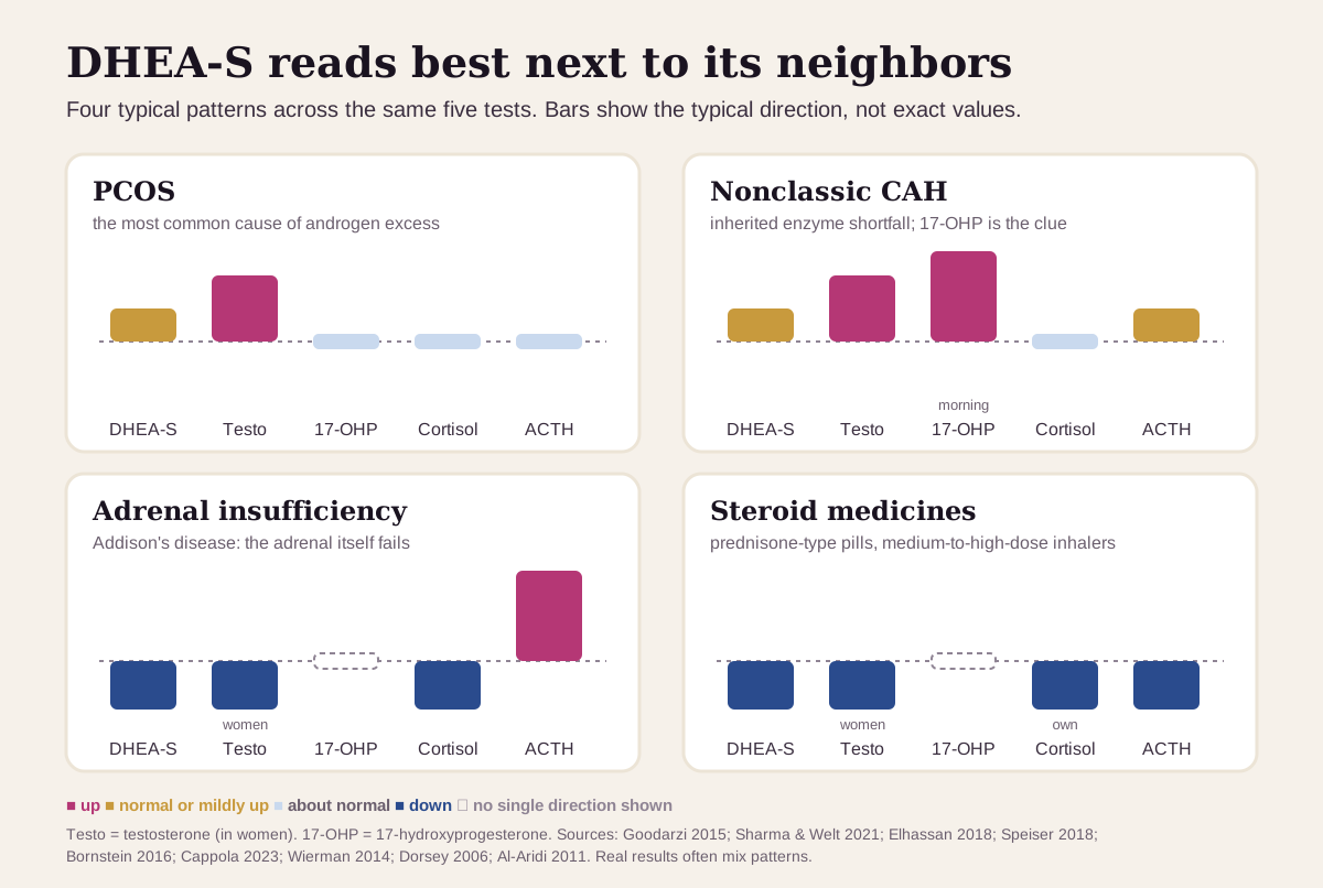
DHEA-S is rarely read alone. The pattern across testosterone, 17-OHP, cortisol and ACTH usually says more than DHEA-S does by itself:75
| If you see… | It often points to… |
|---|---|
| Testosterone up, DHEA-S normal or mildly up, 17-OHP normal | PCOS, the most common cause of excess male hormones in women |
| 17-OHP up (early-morning value over 200 ng/dL), testosterone up | nonclassic CAH: a morning 17-OHP over 500 ng/dL makes the diagnosis |
| DHEA-S over 700 µg/dL, especially after menopause | an adrenal tumor to rule out |
| DHEA-S and cortisol low, ACTH high | adrenal insufficiency (Addison's disease) |
| DHEA-S and your own cortisol low, ACTH low, on steroid medicines | steroids suppressing the adrenal axis |
| DHEA-S low, ACTH low, cortisol not suppressed by dexamethasone, with an adrenal nodule seen on a scan | a cortisol-making adrenal nodule20 |
To see how the adrenals are working, clinicians use morning cortisol, ACTH and a stimulation test. DHEA-S is a quick first screen.195
How fast DHEA-S changes
DHEA-S is steady from hour to hour and falls over decades. It has a long half-life and shows almost no daily rhythm, unlike cortisol, which peaks in the morning.79 You need no special preparation for the test.2
What that means in practice:
- Small changes are noise. In weekly samples from four men, results varied by a coefficient of variation of 19%, with no monthly, seasonal or yearly rhythm.10 A shift of 20% between two tests can be ordinary.
- The decline is slow. Levels fall from your 20s and flatten out in your 50s and 60s, so a steady drift down over years is expected.74
- If you take DHEA, time the test. The Endocrine Society's adrenal guideline measures DHEA-S in the morning, before the daily dose.5
- Trends beat snapshots. Compare results from the same lab, and read each one against the range for your age at the time of the test.4
Going deeper: the adrenal's third product
The adrenal cortex has three layers. The outer layer makes aldosterone, the middle layer makes cortisol, and the inner layer (zona reticularis) makes DHEA and DHEA-S.1 ACTH drives both cortisol and DHEA-S, and cortisol feeds back to turn ACTH down. That loop explains two patterns: steroid medicines lower DHEA-S, and a cortisol-making nodule does too.120
With age, the two outputs split. DHEA-S falls while cortisol holds steady or rises slightly, as the zona reticularis gradually shrinks.1
For adrenal nodules, researchers turned DHEA-S into a DHEA-S ratio:20
DHEA-S ratio = your DHEA-S ÷ the lower limit of your age- and sex-specific range
A ratio of 1.12 or less caught more than 99% of people with mild cortisol excess from an adrenal nodule, with 91.9% specificity.20 It's a good example of DHEA-S working as part of a pattern, read against your own age.
Questions worth asking
- Is my DHEA-S high or low for my age and sex, and by how much?
- What do my testosterone, 17-OHP and SHBG show next to it?
- Should I have an early-morning 17-OHP test to check for nonclassic CAH?
- Could my medicines, inhalers or supplements (including DHEA) be affecting it?
- If it's low, do I need a morning cortisol and ACTH, or a stimulation test?
- When should we repeat it, and what would change the plan?
FAQ
What is a normal DHEA-S level? It depends on your age and sex. MedlinePlus lists 110–510 µg/dL for men and 45–320 µg/dL for women aged 18–29, with lower ranges at older ages.4
What causes a high DHEA-S in women? PCOS is the most common cause. DHEA supplements and nonclassic CAH are others. A level over 700 µg/dL needs a workup for an adrenal tumor.37
Should I take DHEA if my DHEA-S is low? Talk to your clinician first. In a 2-year trial in older adults, DHEA raised DHEA-S but brought no meaningful gains in body composition, strength or quality of life, and the Endocrine Society recommends against routine use in women.1415
When is a high DHEA-S an emergency? A level over 700 µg/dL, fast-growing hair or a deepening voice, or a high level after menopause all need prompt care to rule out a tumor.73
This guide is for education. It can't diagnose you or replace care from a clinician who knows your history.
Footnotes
-
Cappola AR, Auchus RJ, El-Hajj Fuleihan G, et al. Hormones and Aging: An Endocrine Society Scientific Statement. J Clin Endocrinol Metab. 2023;108(8):1835–1874. The source for the adrenal zones, the peak near 25 and fall to childhood levels by 80, women's adrenal share of testosterone, cortisol holding steady with age, and the trial summary on DHEA supplements. PMC ↩ ↩2 ↩3 ↩4 ↩5 ↩6 ↩7 ↩8 ↩9 ↩10 ↩11
-
National Library of Medicine. DHEA Sulfate Test. MedlinePlus. Why the test is ordered, symptoms of high and low levels, and no special preparation. MedlinePlus ↩ ↩2 ↩3 ↩4 ↩5 ↩6 ↩7
-
Elhassan YS, Idkowiak J, Smith K, et al. Causes, Patterns, and Severity of Androgen Excess in 1205 Consecutively Recruited Women. J Clin Endocrinol Metab. 2018;103(3):1214–1223. The DHEA-S severity bands and which diagnoses fell in each. PMC ↩ ↩2 ↩3 ↩4 ↩5 ↩6 ↩7 ↩8 ↩9 ↩10 ↩11 ↩12 ↩13
-
National Library of Medicine. DHEA-sulfate test. MedlinePlus Medical Encyclopedia. Reviewed January 25, 2026. Typical normal ranges by age and sex, causes of high and low results, and glucocorticoid medicines. MedlinePlus ↩ ↩2 ↩3 ↩4 ↩5 ↩6 ↩7 ↩8 ↩9 ↩10 ↩11 ↩12 ↩13
-
Bornstein SR, Allolio B, Arlt W, et al. Diagnosis and Treatment of Primary Adrenal Insufficiency: An Endocrine Society Clinical Practice Guideline. J Clin Endocrinol Metab. 2016;101(2):364–389. Low DHEA-S as an early sign, the cortisol and ACTH criteria, adrenal-crisis symptoms, and the DHEA-S target when women take DHEA. PMC ↩ ↩2 ↩3 ↩4 ↩5 ↩6 ↩7 ↩8 ↩9 ↩10
-
Goodarzi MO, Carmina E, Azziz R. DHEA, DHEAS and PCOS. J Steroid Biochem Mol Biol. 2015;145:213–225. About 20–30% of women with PCOS have adrenal androgen excess, without overt adrenal-axis dysfunction. PubMed ↩ ↩2 ↩3 ↩4
-
Sharma A, Welt CK. Practical Approach to Hyperandrogenism in Women. Med Clin North Am. 2021;105(6):1099–1116. DHEA-S as the marker of adrenal androgens, its long half-life, the 700 µg/dL tumor workup line, and the 17-OHP cutoffs for nonclassic CAH. PMC ↩ ↩2 ↩3 ↩4 ↩5 ↩6 ↩7 ↩8 ↩9 ↩10 ↩11 ↩12 ↩13 ↩14 ↩15
-
National Center for Biotechnology Information. Dehydroepiandrosterone Sulfate (CID 12594). PubChem. Molecular weight 368.5 g/mol, used for the unit conversion. PubChem ↩
-
Turcu AF, Zhao L, Chen X, et al. Circadian rhythms of 11-oxygenated C19 steroids and Δ5-steroid sulfates in healthy men. Eur J Endocrinol. 2021;185(4):K1–K6. DHEA-S showed minimal daily variation. PMC ↩ ↩2 ↩3
-
Orentreich N, Brind JL, Rizer RL, Vogelman JH. Age changes and sex differences in serum dehydroepiandrosterone sulfate concentrations throughout adulthood. J Clin Endocrinol Metab. 1984;59(3):551–555. 981 men and 481 women; peak and after-70 averages; 19% week-to-week variation. PubMed ↩ ↩2 ↩3 ↩4
-
Optimal DX. Hormone Biomarkers: DHEA-S. Updated April 14, 2025. States an optimal DHEA-S of 350–690 µg/dL for men and 275–390 µg/dL for women. A commercial source, listed as what the optimizer community uses. optimaldx.com ↩ ↩2 ↩3
-
Guilliams TG. The Role of Stress and the HPA Axis in Chronic Disease Management. Point Institute; 2015. ISBN 978-0985615826. The functional-medicine textbook the Libby app's marker catalog cites for its age- and sex-specific optimal DHEA-S ranges. ↩ ↩2 ↩3 ↩4 ↩5
-
Ohlsson C, Labrie F, Barrett-Connor E, et al. Low serum levels of dehydroepiandrosterone sulfate predict all-cause and cardiovascular mortality in elderly Swedish men. J Clin Endocrinol Metab. 2010;95(9):4406–4414. MrOS Sweden, 2,644 men aged 69–81, 4.5 years of follow-up. PubMed ↩ ↩2 ↩3
-
Nair KS, Rizza RA, O'Brien P, et al. DHEA in elderly women and DHEA or testosterone in elderly men. N Engl J Med. 2006;355(16):1647–1659. Two-year randomized, placebo-controlled trial in 87 men and 57 women with low DHEA-S. PubMed ↩ ↩2 ↩3
-
Wierman ME, Arlt W, Basson R, et al. Androgen therapy in women: a reappraisal: an Endocrine Society clinical practice guideline. J Clin Endocrinol Metab. 2014;99(10):3489–3510. Recommends against routine DHEA in women; lists hypopituitarism, adrenal insufficiency and glucocorticoid use as causes of low androgens. PubMed ↩ ↩2 ↩3
-
Speiser PW, Arlt W, Auchus RJ, et al. Congenital Adrenal Hyperplasia Due to Steroid 21-Hydroxylase Deficiency: An Endocrine Society Clinical Practice Guideline. J Clin Endocrinol Metab. 2018;103(11):4043–4088. Nonclassic CAH frequency (about 1 in 200 in the U.S.) and its mild effect on cortisol. PMC ↩ ↩2
-
d'Alva CB, Abiven-Lepage G, Viallon V, et al. Sex steroids in androgen-secreting adrenocortical tumors: clinical and hormonal features in comparison with non-tumoral causes of androgen excess. Eur J Endocrinol. 2008;159(5):641–647. Tumors are 0.2% or less of androgen excess; DHEA-S high in 82% of them. PubMed ↩
-
Dorsey MJ, Cohen LE, Phipatanakul W, et al. Assessment of adrenal suppression in children with asthma treated with inhaled corticosteroids: use of dehydroepiandrosterone sulfate as a screening test. Ann Allergy Asthma Immunol. 2006;97(2):182–186. PubMed ↩ ↩2
-
Al-Aridi R, Abdelmannan D, Arafah BM. Biochemical diagnosis of adrenal insufficiency: the added value of dehydroepiandrosterone sulfate measurements. Endocr Pract. 2011;17(2):261–270. A normal age- and sex-adjusted DHEA-S practically rules out adrenal insufficiency. PubMed ↩ ↩2 ↩3
-
Dennedy MC, Annamalai AK, Prankerd-Smith O, et al. Low DHEAS: A Sensitive and Specific Test for the Detection of Subclinical Hypercortisolism in Adrenal Incidentalomas. J Clin Endocrinol Metab. 2017;102(3):786–792. 185 patients; a DHEA-S ratio of 1.12 was more than 99% sensitive and 91.9% specific; ACTH under 10 pg/mL in every case. PubMed ↩ ↩2 ↩3 ↩4 ↩5
Educational content, not medical advice.Libby is a personal record tool, not a medical service — it doesn't diagnose, treat, or prescribe. Reference ranges vary by lab and by person. Talk to a qualified healthcare professional about your results.
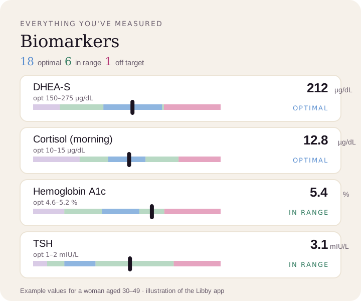
Compare your results to optimal ranges.
Upload a lab PDF or a photo of your report. Libby puts every marker on your lab's range and the optimal range, and tracks your trends over time.
View your DHEA-S ›