Rheumatoid factor (RF): normal range and what high means
Rheumatoid factor is an antibody tied to rheumatoid arthritis. What a normal result is, what a positive one usually means, and how to read it with anti-CCP.
Rheumatoid factor (RF) is an antibody that sticks to your own antibodies, and it's the classic blood test for rheumatoid arthritis. In people with rheumatoid arthritis (RA), RF is positive about 7 times in 10, so a positive result supports that diagnosis when joints are swollen.1 RF also shows up in Sjögren's disease, hepatitis C and other long-lasting infections, and in some healthy people, more often with age.23
The short version
- Normal: below your lab's cutoff, most often under 14 IU/mL (Labcorp and ARUP). Cutoffs on different test kits run from 12 to 30 IU/mL.456
- Positive points toward RA when joints are swollen, and it's common elsewhere too. Pooled across 50 studies, RF was positive in 69% of people with RA and in 15% of comparison patients without it.1
- Size matters. Above 3 times your lab's cutoff (over 42 IU/mL when the cutoff is 14) counts as "high-positive" in the RA classification criteria.7 In 9,712 Danish adults, an RF over 100 IU/mL came with 26 times the long-term risk of developing RA.8
- No RF level is an emergency. A hot, swollen, very painful joint with fever is: get care the same day.9
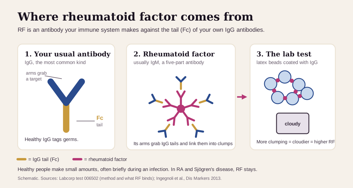
What rheumatoid factor is
Antibodies are the proteins your immune system uses to tag germs. The most common kind, IgG, has two arms that grab a target and a tail called the Fc region. Rheumatoid factor is an antibody against that tail. It's usually an IgM antibody, though IgG and IgA versions exist.42 (More on the types at the end.)
Healthy immune systems make some RF. During an infection, B cells can pump out low-affinity IgM RF that helps clear clumps of antibodies and germs, and that RF usually fades afterward.2 In RA and Sjögren's disease, the RF-making cells keep going, and Sjögren's disease produces some of the highest levels seen.2
The test is a clumping test. At Labcorp, for example, your serum is mixed with tiny latex beads coated with human IgG. RF links the beads together, and the analyzer reads how cloudy the sample turns (latex immunoturbidimetry).4 More clumping means a higher number.
Clinicians order RF mainly when joints are painful, stiff or swollen, usually alongside anti-CCP, a more RA-specific antibody, and CRP or ESR, which measure inflammation.11107 It's also part of the workup for Sjögren's disease, an autoimmune condition that dries the eyes and mouth.1012
What's a normal rheumatoid factor?
Normal is below your own lab's cutoff. For the big U.S. reference labs that's under 14 IU/mL: Labcorp prints <14.0 and ARUP prints 0–14.45 MedlinePlus gives under 15 IU/mL, or a titer under 1:80, as typical.10
Different test kits draw the line in different places. A 2022 study ran nine RF tests on the same 1,471 samples. The manufacturers' cutoffs ranged from 12 to 30 IU/mL, and the international RF reference serum read negative on some kits and positive on others.6
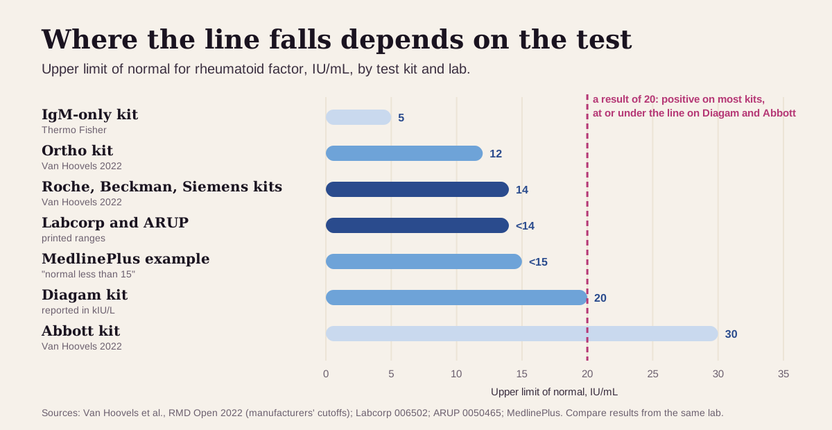
So a result of 20 can be positive at one lab and negative at another. Compare a new result with earlier ones from the same lab.
Who tests positive? In U.S. national surveys, a positive RF rose with age from 0.7% of adults aged 18–24 to 5.8% of those over 60 (1960–62), and 6.5% of adults over 60 tested positive in 1988–94. Men and women were about equally likely to test positive.3 A review puts the share at 10–25% of healthy 70-year-olds.2 One large Danish study is the exception: it found similar RF levels from age 20 to 100.8
"Optimal" rheumatoid factor: what health optimizers aim for
For RF the goal is simple: lower is better, and the strictest aim for undetectable.13 Here's the ladder, strictest first. These limits come from different tests, so read them as a rough order rather than one ruler.
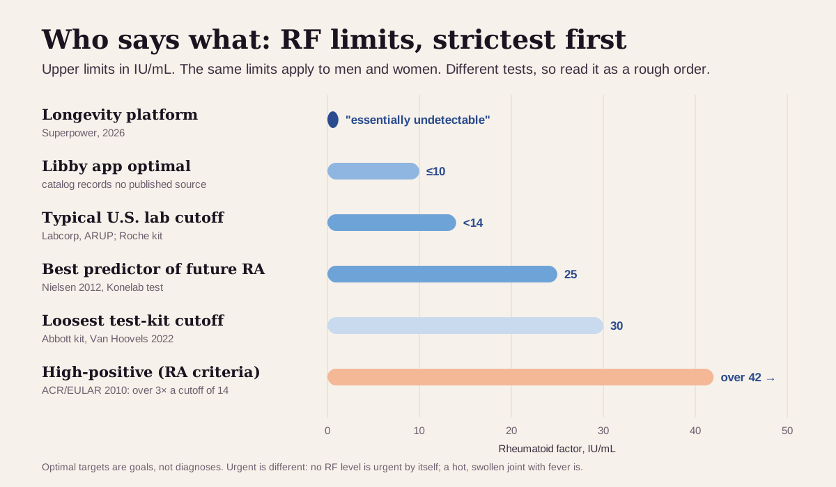
| Target | Upper limit (IU/mL) | Who uses it |
|---|---|---|
| Strictest (longevity platform) | "essentially undetectable" | Superpower's published optimal13 |
| Libby app optimal | 10 or less | The optimal range shown in the Libby app, the same for men and women. The app's catalog records no published source for this band. On the Roche test, 10 IU/mL is the bottom of the measuring range, so there this band sits at the floor of what the test can read6 |
| Typical U.S. lab cutoff | under 14 | Labcorp and ARUP, and the Roche, Beckman Coulter and Siemens kits456 |
| Research: best predictor of future RA | 25 | The level that best separated people who later developed RA in the Copenhagen City Heart Study (a different test, measured on a Konelab analyzer)8 |
| Loosest test-kit cutoff | 30 | Abbott's kit6 |
| Classification "high-positive" | over 3× your lab's cutoff (over 42 at a cutoff of 14) | The 2010 ACR/EULAR rheumatoid arthritis criteria, which give it 3 of the 10 points7 |
| Urgent | none | No RF level is urgent on its own. Urgency comes from a hot, swollen, very painful joint with fever9 |
Why the low targets aren't just a trend:
- In the Copenhagen City Heart Study, 9,712 adults without RA gave blood in 1981–83 and were followed for up to 28 years. Compared with an RF under 25 IU/mL, the risk of developing RA was 3.6 times higher at 25–50, 6.0 times at 50–100 and 26 times above 100. Over the first 10 years, those figures were 6.0, 14 and 39.8
- In absolute terms, the highest 10-year risk was 32%, in women aged 50–69 who smoked and had an RF over 100. The lowest was 0.1%, in men over 70 with an RF under 25.8
- RF often shows up years before joint symptoms. Among 79 blood donors who later developed RA, 49% already had RF, anti-CCP or both a median of 4.5 years before their symptoms began. Only 1.1% of control samples were RF-positive.14
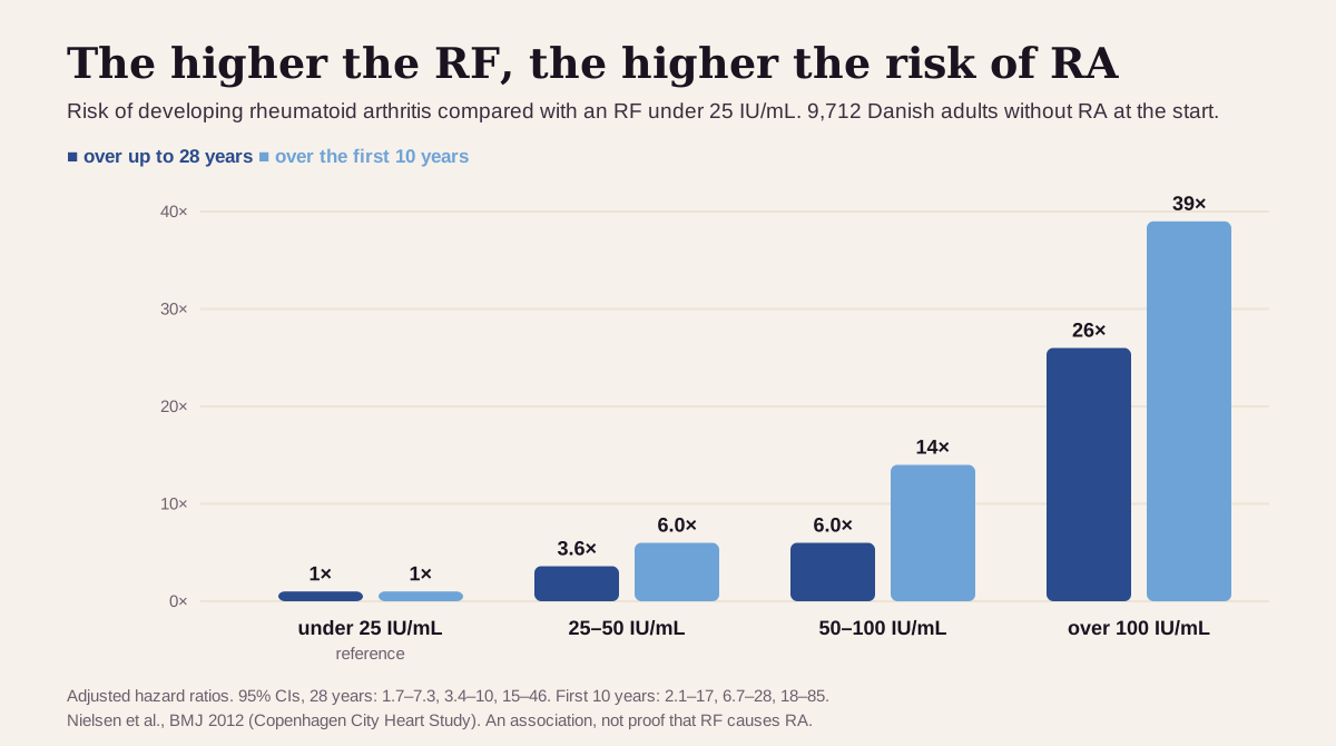
Keep "optimal" in perspective: RF is a risk marker, and the number itself is rarely what clinicians treat. Immunosuppressive treatment can lower RF, yet RF tracks disease activity poorly,2 and in 640 people with early arthritis, repeating RF after 5 years added nothing to the first result for predicting joint damage.15 Even in the highest-risk Danish group, about two in three stayed free of RA for 10 years. The authors called their smoking finding one more reason for women with a raised RF to quit.8
What a positive rheumatoid factor usually means
Start with the size. The 2010 RA classification criteria from the American College of Rheumatology and the European League Against Rheumatism (ACR/EULAR) sort RF into three bands against your lab's upper limit of normal (ULN): negative at or below it, low-positive up to 3 times it, and high-positive above that.7
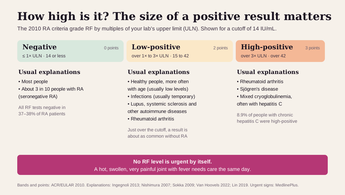
The usual suspects:
- Rheumatoid arthritis. RA affects about 1% of people.6 Pooled across studies, RF is positive in 69% of them.1 RA usually brings joint pain and stiffness that's worse in the morning, in the same joints on both sides of the body.11 Among people with RA, a high RF goes with more severe disease on average.4
- Getting older. About 1 in 15 U.S. adults over 60 tests positive, and the RF found in healthy people is usually low to moderate.32
- Sjögren's disease. Of 10,500 people at diagnosis, 48.6% were RF-positive.18 Along with mixed cryoglobulinemia (RF-antibody complexes that settle out of the blood when it's cooled),19 it produces the highest RF levels.2
- Hepatitis C and other long infections. In 271 people with chronic hepatitis C and no arthritis, 47.2% had a positive RF and 8.9% a high-positive one.17 A review names hepatitis C as a leading cause of raised RF and advises a hepatitis C antibody test for anyone with one. Endocarditis, an infection of the heart valves (40%), and tuberculosis (15%) raise it too.2
- Other autoimmune diseases: mixed connective tissue disease (50–60%), lupus (15–35%), systemic sclerosis (20–30%) and dermatomyositis (20%).2
- Less common: primary biliary cholangitis (a slow autoimmune liver disease), chronic sarcoidosis, some cancers such as leukemia and myeloma, and a run of recent vaccinations.210
A negative RF with swollen joints
About a third of people with RA test RF-negative. In two clinics that tracked RA patients from 1980 to 2004, every RF test was negative in 37–38% of them.16 The ACR/EULAR criteria can still reach their 6-point threshold without any antibody, through the number of swollen joints, a raised CRP or ESR, and symptoms lasting 6 weeks or more.7 That's called seronegative RA.
Get care the same day for a joint that quickly turns red, swollen and very painful, especially with a fever. That can be a joint infection (septic arthritis), and RA raises the risk. Delayed treatment can damage the joint permanently.9
Rheumatoid factor reads best next to its neighbors
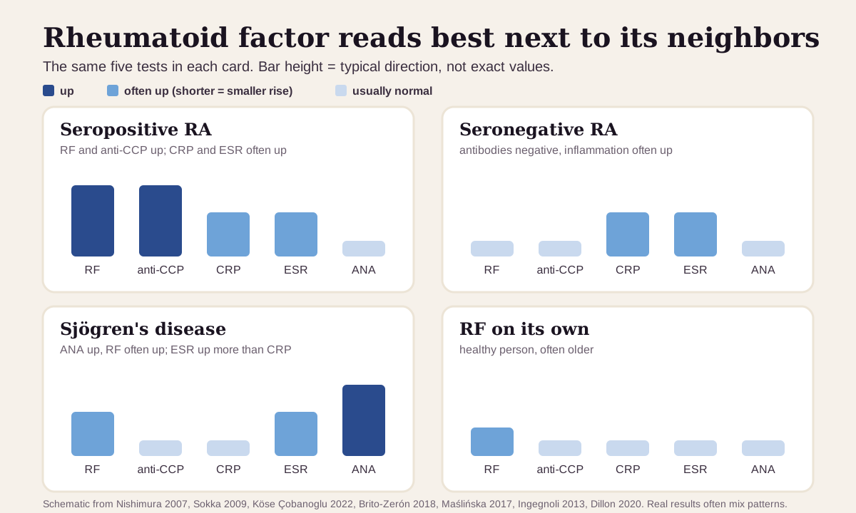
RF on its own points in several directions. Four other common tests sort out most of them: anti-CCP (a more RA-specific antibody), CRP and ESR (inflammation), and ANA (a broad autoimmune screen).120
| If you see… | It often points to… |
|---|---|
| RF and anti-CCP both positive, often with a high CRP or ESR | the classic seropositive RA pattern. Anti-CCP is the more specific of the two: positive in 5% of comparison patients without RA, versus 15% for RF1 |
| RF and anti-CCP negative, CRP or ESR up, swollen joints | seronegative RA is still possible: every RF test was negative in 37–38% of RA patients at two clinics (1980–2004),16 and the RA criteria can be met with both antibodies negative7 |
| RF and ANA positive, anti-CCP negative, ESR up more than CRP | a Sjögren's pattern: at diagnosis, 79% have ANA and 49% RF.18 Anti-CCP turns up in only 6–13%, and CRP and ESR run lower than in RA2012 |
| RF positive, anti-CCP negative, a hepatitis C risk or known infection | chronic hepatitis C: RF in 47% versus anti-CCP in 1.1%17 |
| RF low-positive and everything else normal, no swollen joints | RF in a healthy person, found more often with age in U.S. surveys.23 The risk of later RA rises with the level8 |
RF and anti-CCP work as a pair. Pooled across studies, anti-CCP was about as sensitive as IgM RF (67% versus 69%) but more specific (95% versus 85%). A positive anti-CCP multiplies the odds of RA about 12-fold, versus about 5-fold for RF.1 Our anti-CCP guide covers that test in detail.
How fast rheumatoid factor changes
RF builds up before RA starts. In Swedish blood donors who later developed RA, IgM RF was already positive in 15% more than 1.5 years before their first symptoms, and in 30% during the last 1.5 years.21 In the Danish study, the higher the RF, the sooner RA came: a median of 15 years after the blood test when RF was under 25, and 7 years when it was over 50.8
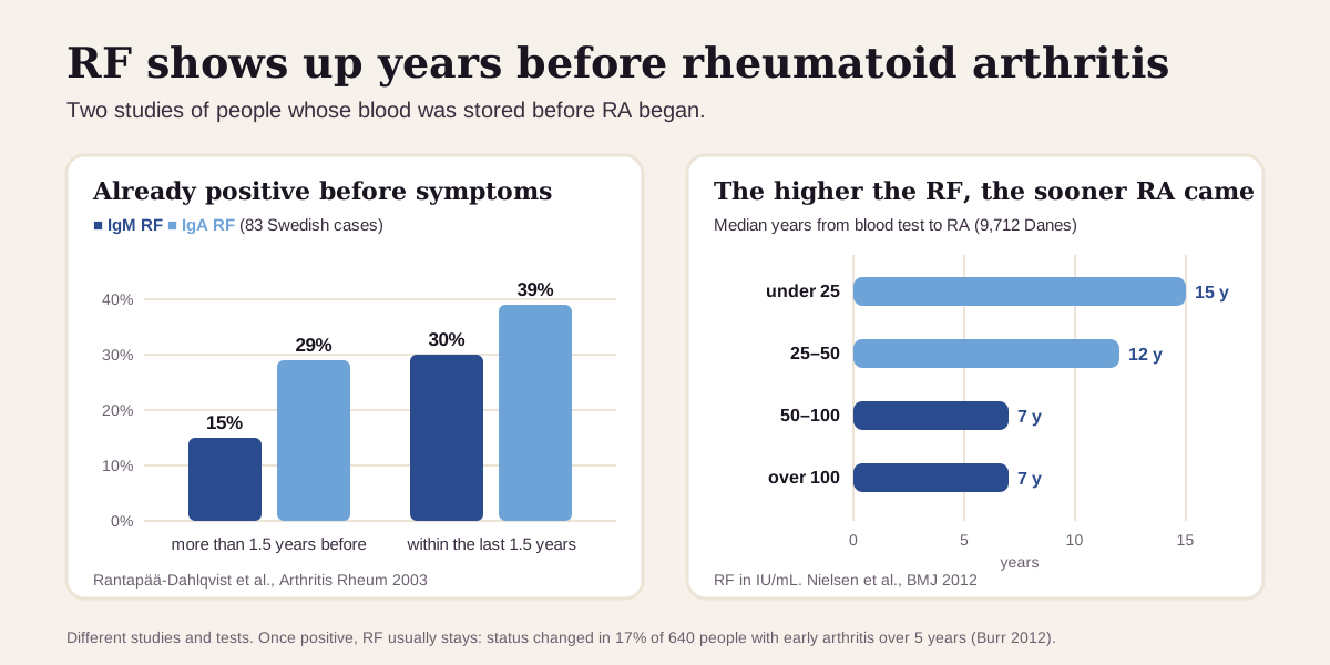
What that means in practice:
- RF moves slowly, and one result usually stands. Among 640 people with early arthritis, RF status (positive or negative) changed in 17% over 5 years, and the second test added nothing for predicting outcome.15
- RF from an infection is usually temporary.2 If a low-positive result came during or soon after an illness, mention that to whoever reads it.
- No preparation is needed at most labs, though ARUP prefers a fasting sample.115 Very fatty (lipemic) samples can give a false positive on Labcorp's test.4
- Treatment can lower RF, yet the number tracks RA activity poorly.2
Going deeper: what the number carries
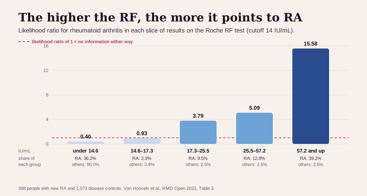
A bigger number carries more weight. The 2022 multicenter study worked out a likelihood ratio for each slice of RF results. On the Roche test (cutoff 14), a result just over the line, 14.6–17.3 IU/mL, had a likelihood ratio of 0.93: about as common in people without RA as in people with it. Above 57.2 it was 15.6, and 39% of people with RA landed there.6
RF comes in types. Routine tests measure total RF, which is mostly IgM.4 Specialty labs can measure IgM, IgA and IgG RF separately. All three turn up together in up to 52% of people with RA, versus fewer than 5% of people with other connective-tissue diseases.2 Before RA, they tend to appear in order: IgM first, then IgA, then IgG.2 In the Swedish donor study, IgA RF and anti-CCP were the two significant predictors of RA.21
RF can throw off other tests. Because it binds antibodies, RF can interfere with immunoassays for other things, including tests for rubella and hepatitis C antibodies, some thyroid tests, and the tumor marker CA 19-9.2 If an odd result shows up alongside a high RF, ask the lab about it.
How the criteria use it. The 2010 ACR/EULAR system applies to someone with at least one swollen joint and no better explanation, and it needs 6 of 10 points. Serology supplies up to 3: 0 for negative RF and anti-CCP, 2 for low-positive, 3 for high-positive. Joints supply up to 5, an abnormal CRP or ESR 1, and symptoms lasting 6 weeks or more 1.7 These are classification rules, built to recognize RA early and consistently in research.
Questions worth asking
- Is my RF low-positive or high-positive by my lab's cutoff?
- Have I had an anti-CCP test, and what did it show?
- Do my CRP and ESR suggest active inflammation?
- Should I be tested for hepatitis C?
- Could dry eyes or a dry mouth point to Sjögren's disease?
- If I have no joint symptoms, what would prompt a recheck?
FAQ
What is a normal rheumatoid factor level? Below your lab's cutoff, most often under 14 IU/mL. Test kits use cutoffs from 12 to 30 IU/mL, so check the range on your own report.46
Does a positive RF mean I have rheumatoid arthritis? Only together with the right symptoms. RF is positive in about 69% of people with RA, and also in 15% of comparison patients without RA, about half of people with Sjögren's disease or chronic hepatitis C, and some healthy older adults.118173 A high-positive RF plus a positive anti-CCP and swollen joints makes RA much more likely.7
Can you have rheumatoid arthritis with a negative RF? Yes. At two clinics followed from 1980 to 2004, every RF test was negative in 37–38% of people with RA. That's seronegative RA, recognized from the joints, inflammation markers and how long symptoms have lasted.167
Should RF be retested? Usually only when the question changes, such as new joint symptoms. RF status changed in 17% of people with early arthritis over 5 years, and repeating it added nothing for predicting outcome.15
This guide is for education. It can't diagnose you or replace care from a clinician who knows your history.
Footnotes
-
Nishimura K, Sugiyama D, Kogata Y, Tsuji G, et al. Meta-analysis: diagnostic accuracy of anti-cyclic citrullinated peptide antibody and rheumatoid factor for rheumatoid arthritis. Ann Intern Med. 2007;146(11):797–808. Pooled 50 RF and 37 anti-CCP studies: IgM RF sensitivity 69%, specificity 85%, positive likelihood ratio 4.86; anti-CCP 67%, 95%, 12.46. PubMed ↩ ↩2 ↩3 ↩4 ↩5 ↩6 ↩7 ↩8 ↩9
-
Ingegnoli F, Castelli R, Gualtierotti R. Rheumatoid factors: clinical applications. Dis Markers. 2013;35(6):727–734. Review of what RF is, its isotypes, how often it appears in each condition (Table 1), and its interference with other tests. PMC ↩ ↩2 ↩3 ↩4 ↩5 ↩6 ↩7 ↩8 ↩9 ↩10 ↩11 ↩12 ↩13 ↩14 ↩15 ↩16 ↩17 ↩18 ↩19 ↩20
-
Dillon CF, Weisman MH, Miller FW. Population-based estimates of humoral autoimmunity from the U.S. National Health and Nutrition Examination Surveys, 1960–2014. PLoS One. 2020;15(1):e0226516. U.S. national survey RF prevalence by age and sex. PMC ↩ ↩2 ↩3 ↩4 ↩5 ↩6
-
Labcorp, a commercial lab. Rheumatoid Factor (RF), test 006502. Method (latex immunoturbidimetry), reference range (<14.0 IU/mL), and notes on what RF is, false positives from lipemic samples and RF in healthy people. labcorp.com ↩ ↩2 ↩3 ↩4 ↩5 ↩6 ↩7 ↩8 ↩9 ↩10 ↩11 ↩12
-
ARUP Laboratories, a commercial lab. Rheumatoid Factor, test 0050465. Reference interval 0–14 IU/mL by immunoturbidimetry; fasting specimen preferred. aruplab.com ↩ ↩2 ↩3 ↩4 ↩5 ↩6
-
Van Hoovels L, Vander Cruyssen B, Sieghart D, Bonroy C, et al. Multicentre study to improve clinical interpretation of rheumatoid factor and anti-citrullinated protein/peptide antibodies test results. RMD Open. 2022;8(1):e002099. Nine RF tests from different manufacturers on samples from 398 people with newly diagnosed RA and 1,073 disease controls: manufacturers' cutoffs and result-specific likelihood ratios. PMC ↩ ↩2 ↩3 ↩4 ↩5 ↩6 ↩7 ↩8 ↩9 ↩10 ↩11
-
Aletaha D, Neogi T, Silman AJ, Funovits J, et al. 2010 rheumatoid arthritis classification criteria: an American College of Rheumatology/European League Against Rheumatism collaborative initiative. Ann Rheum Dis. 2010;69(9):1580–1588. The source for the 6-of-10 rule, the scoring domains and the negative / low-positive (over ULN, up to 3× ULN) / high-positive (over 3× ULN) definitions, as set out in EULAR's official criteria slides. PubMed ↩ ↩2 ↩3 ↩4 ↩5 ↩6 ↩7 ↩8 ↩9 ↩10 ↩11
-
Nielsen SF, Bojesen SE, Schnohr P, Nordestgaard BG. Elevated rheumatoid factor and long term risk of rheumatoid arthritis: a prospective cohort study. BMJ. 2012;345:e5244. 9,712 adults in the Copenhagen City Heart Study, followed up to 28 years. PMC ↩ ↩2 ↩3 ↩4 ↩5 ↩6 ↩7 ↩8 ↩9 ↩10 ↩11
-
National Library of Medicine. Septic arthritis. MedlinePlus Medical Encyclopedia. Symptoms of a joint infection and why prompt treatment matters. MedlinePlus ↩ ↩2 ↩3
-
National Library of Medicine. Rheumatoid factor (RF). MedlinePlus Medical Encyclopedia. Typical normal values (under 15 IU/mL, or titer under 1:80) and conditions with high RF. MedlinePlus ↩ ↩2 ↩3 ↩4 ↩5 ↩6
-
National Library of Medicine. Rheumatoid Factor (RF) Test. MedlinePlus. What the test is for, RA symptoms, other conditions with RF, and preparation. MedlinePlus ↩ ↩2 ↩3
-
Maślińska M, Mańczak M, Wojciechowska B, Kwiatkowska B. The prevalence of ANA antibodies, anticentromere antibodies, and anti-cyclic citrullinated peptide antibodies in patients with primary Sjögren's syndrome compared to patients with dryness symptoms without primary Sjögren's syndrome confirmation. Reumatologia. 2017;55(3):113–119. ANA in 88% and anti-CCP in 13% of 75 people with Sjögren's disease; raised ESR among the features that set it apart. PMC ↩ ↩2 ↩3
-
Superpower. Rheumatoid Factor. September 1, 2026. States that "optimal" RF "is essentially undetectable." A commercial source, listed as what the optimizer community uses. superpower.com ↩ ↩2 ↩3
-
Nielen MM, van Schaardenburg D, Reesink HW, van de Stadt RJ, et al. Specific autoantibodies precede the symptoms of rheumatoid arthritis: a study of serial measurements in blood donors. Arthritis Rheum. 2004;50(2):380–386. PubMed ↩
-
Burr ML, Viatte S, Bukhari M, Plant D, et al. Long-term stability of anti-cyclic citrullinated peptide antibody status in patients with early inflammatory polyarthritis. Arthritis Res Ther. 2012;14(3):R109. 640 people in the Norfolk Arthritis Register tested at baseline and 5 years. PMC ↩ ↩2 ↩3
-
Sokka T, Pincus T. Erythrocyte sedimentation rate, C-reactive protein, or rheumatoid factor are normal at presentation in 35%–45% of patients with rheumatoid arthritis seen between 1980 and 2004: analyses from Finland and the United States. J Rheumatol. 2009;36(7):1387–1390. PubMed ↩ ↩2 ↩3 ↩4 ↩5
-
Lin KM, Chen WM, Tung SY, Wei KL, et al. Prevalence and predictive value of high-positive rheumatoid factor and anti-citrullinated protein antibody levels in nonarthritic patients with chronic hepatitis C infection. Int J Rheum Dis. 2019;22(1):116–120. 271 people with chronic hepatitis C: RF 47.2%, high-positive RF 8.9%, anti-CCP 1.1%. PubMed ↩ ↩2 ↩3 ↩4
-
Brito-Zerón P, Acar-Denizli N, Ng WF, Zeher M, et al. How immunological profile drives clinical phenotype of primary Sjögren's syndrome at diagnosis: analysis of 10,500 patients (Sjögren Big Data Project). Clin Exp Rheumatol. 2018;36 Suppl 112(3):102–112. At diagnosis: ANA 79.3%, anti-Ro 73.2%, RF 48.6%. PubMed ↩ ↩2 ↩3 ↩4
-
Gorevic PD. Rheumatoid factor, complement, and mixed cryoglobulinemia. Clin Dev Immunol. 2012;2012:439018. Defines mixed cryoglobulins as "cold-precipitable rheumatoid factors." PMC ↩
-
Köse Çobanoglu R, Şentürk T. The role of albumin-to-globulin ratio in undifferentiated arthritis: rheumatoid arthritis versus primary Sjögren syndrome. Arch Rheumatol. 2022;37(2):245–251. 96 people with RA and 81 with Sjögren's disease: ESR and CRP higher in RA; RF and ANA more common in Sjögren's; anti-CCP 6.2% in Sjögren's. PMC ↩ ↩2 ↩3
-
Rantapää-Dahlqvist S, de Jong BA, Berglin E, Hallmans G, et al. Antibodies against cyclic citrullinated peptide and IgA rheumatoid factor predict the development of rheumatoid arthritis. Arthritis Rheum. 2003;48(10):2741–2749. 83 people in northern Sweden whose blood was stored before RA began. PubMed ↩ ↩2 ↩3
Educational content, not medical advice. Libby is a personal record tool, not a medical service — it doesn't diagnose, treat, or prescribe. Reference ranges vary by lab and by person. Talk to a qualified healthcare professional about your results.
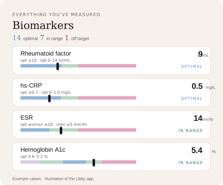
Compare your results to optimal ranges.
Upload a lab PDF or a photo of your report. Libby puts every marker on your lab's range and the optimal range, and tracks your trends over time.
View your rheumatoid factor ›