Plasma free hemoglobin: normal range and high causes
Plasma free hemoglobin measures hemoglobin loose in your blood. What's normal, how a hard blood draw fakes a high result, and when it's urgent.
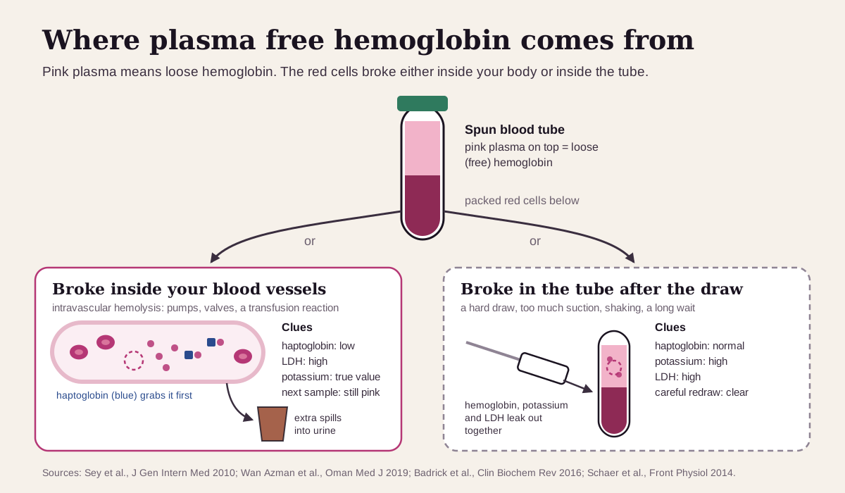
Plasma free hemoglobin measures hemoglobin floating loose in the liquid part of your blood, outside the red cells. Hemoglobin normally stays packed inside red blood cells.1 When red cells burst inside your blood vessels, their hemoglobin spills into the plasma, and this test picks it up.23 The catch: red cells that break in the tube after a hard blood draw release hemoglobin too, and that looks the same on the report.34
The short version
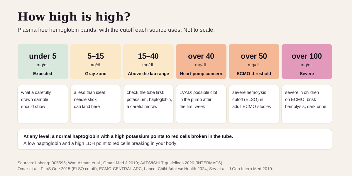
- Normal: very low. Labcorp's range is under 5 mg/dL when the blood is drawn carefully, and it reads 5–15 mg/dL with caution because a rough draw can push a result there. ARUP's range is 0–9.7 mg/dL.35
- The biggest trap is the tube. Red cells broken during a difficult draw or rough handling raise the result. Haptoglobin and potassium tell the two apart: in the tube, potassium rises and haptoglobin stays normal; in the body, haptoglobin falls.46
- Where it's used: heart-lung bypass and ECMO, heart pumps (LVADs) and artificial heart valves, suspected transfusion reactions, and other conditions where red cells break inside the vessels, such as sickle cell disease and PNH.3789
- Urgent: heart-pump teams worry above 40 mg/dL, and ECMO teams use 50 mg/dL as a hemolysis threshold. Red, brown or cola-colored urine with a high result needs same-day care.7102
What plasma free hemoglobin is
Hemoglobin is the iron-rich protein that carries oxygen. It lives inside red blood cells, and old red cells are normally broken down in the spleen, liver and bone marrow, with the hemoglobin kept inside cells the whole time.1 When red cells burst inside the blood vessels instead, a doctor calls it intravascular hemolysis, and the hemoglobin lands in the plasma.2
Your body has a cleanup crew for that. Haptoglobin, a protein made by the liver, grabs loose hemoglobin first, and the pair is quickly cleared by white blood cells called macrophages.411 Only when haptoglobin is used up does loose hemoglobin build up in the plasma, and with brisk breakdown it spills into the urine (hemoglobinuria).2 (Our haptoglobin guide covers that half of the story.)
So plasma free hemoglobin is a "right now" test for red cells breaking inside your vessels. It's mostly a hospital test. Labcorp lists its uses as checking for intravascular hemolysis, including a mismatched (ABO-incompatible) transfusion, red cells damaged mechanically ("traumatic hemolysis"), falciparum malaria, burns, and march hemoglobinuria.3
What's a normal plasma free hemoglobin?
Normal is close to zero, and each lab sets its own upper limit:
- Labcorp: under 5 mg/dL "if conditions are optimal." Results of 5–15 mg/dL "should be interpreted with caution," because a less than ideal needle stick can push results into that range.3
- ARUP Laboratories: 0.0–9.7 mg/dL.5
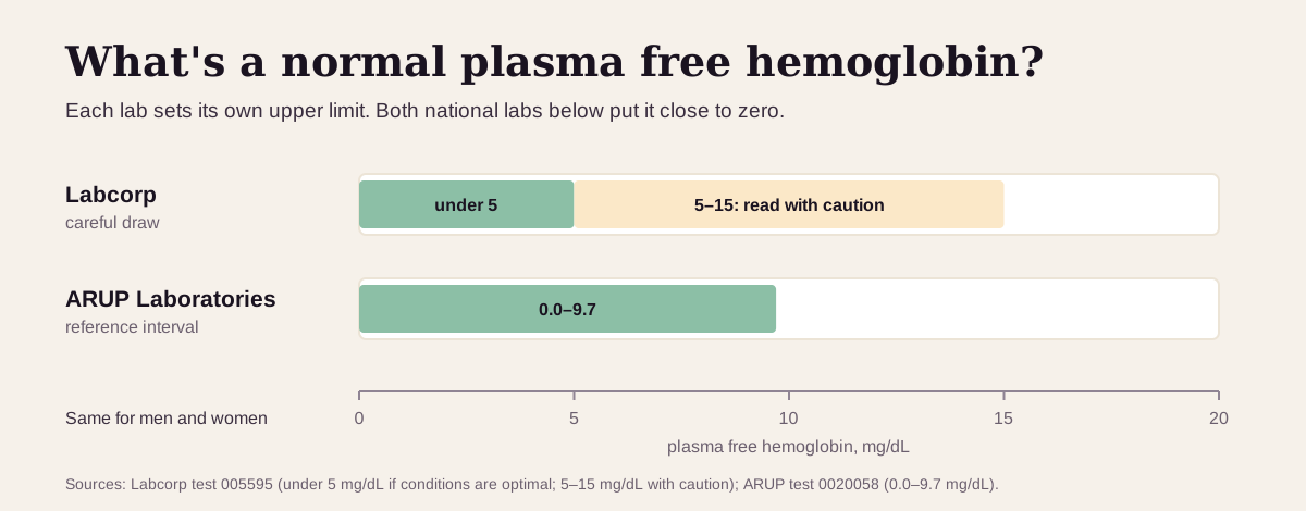
The ranges differ partly because the methods differ. Labcorp measures hemoglobin by the color change it causes in a chemical reaction; ARUP measures it by spectrophotometry, how much light the plasma absorbs.35 That's why a trend is best read from one lab.
The way the blood is drawn matters a lot for this test. Labcorp's instructions call for a large 18-gauge needle, a light tourniquet, throwing away the first 3 mL of blood, mixing the tube gently only three to five times, and spinning the plasma off right away.3 ARUP asks for the plasma to be separated within 2 hours, because red cells left sitting in the tube raise the result.5 Both labs want a green-top heparin tube.35
The same cutoffs apply to men and women.
"Optimal" plasma free hemoglobin: what the thresholds are
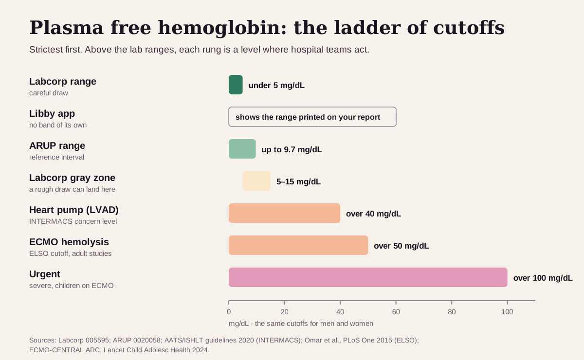
For most markers, health optimizers chase a number inside the normal range. Plasma free hemoglobin works differently: the goal is as low as the method can measure, and the rungs above normal are action thresholds that hospital teams use. Here's the ladder, strictest first:
| Target | Cutoff | Who uses it |
|---|---|---|
| Strictest lab range | under 5 mg/dL | Labcorp, "if conditions are optimal"3 |
| Libby app | the range printed on your report | Libby shows plasma free hemoglobin as your lab reports it ("Reported as-is"), with your lab's range |
| Other national lab | 0.0–9.7 mg/dL | ARUP Laboratories' reference interval5 |
| Gray zone | 5–15 mg/dL | Labcorp: read with caution, since a rough draw can land here3 |
| Heart pump (LVAD) concern | over 40 mg/dL | The U.S. heart-pump registry (INTERMACS) definition of hemolysis; after the first week it suggests a clot in the pump7 |
| ECMO hemolysis threshold | over 50 mg/dL | The Extracorporeal Life Support Organization's cutoff for severe hemolysis, as used in adult ECMO studies10 |
| Urgent | over 100 mg/dL, or any high result with red or brown urine | "Severe hemolysis" in the international definitions for children on ECMO;12 red urine from hemolysis needs urgent workup2 |
Why the thresholds exist:
- In 154 adults on ECMO, a plasma free hemoglobin over 50 mg/dL one day after starting turned up in 15.5% of those who died versus 3.9% of survivors, and it carried about 3.4 times the risk of dying in hospital after accounting for other factors.10
- In 67 people having heart surgery, plasma free hemoglobin rose and haptoglobin fell during surgery on the heart-lung machine, but not in off-pump surgery. The 9 people (13.4%) who developed kidney injury already had higher free hemoglobin during the operation.13
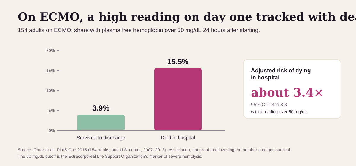
Keep "optimal" in perspective: these are hospital action levels, not goals for a healthy person to beat. A normal result on a carefully drawn sample is the target. Loose hemoglobin matters because it soaks up nitric oxide, the signal that relaxes blood vessels and other smooth muscle, which is one likely way it harms the kidneys and gut (more below).913
What a high plasma free hemoglobin usually means
First question: did the red cells break in you, or in the tube? Hemolyzed samples are the most common sample problem labs see: up to 3.3% of all routine samples, and 40–70% of all samples labs flag as unusable.144
The usual suspect: red cells broken in the tube
Red cells break outside the body from the wrong needle size, a tourniquet left on too long, too much suction, a difficult draw, a wrongly filled tube, mixing the tube the wrong way, jarring transport such as a hospital's pneumatic-tube system, heat or cold, and letting the tube sit before it's spun.4
How labs tell:
- Potassium, LDH and AST rise together with the hemoglobin, because they all leak out of the broken cells in the tube. With breakdown inside the body, the potassium result is the true one.4
- Haptoglobin stays normal. Breakage in the tube leaves your haptoglobin untouched; only breakdown inside your body uses it up. Labs call it the most useful test for telling the two apart.4
- A second, careful sample is clear. If the first tube is pink and the next one isn't, the tube is the likely cause. If sample after sample is pink, labs consider a real problem, especially after a transfusion, on a bypass pump, on dialysis or ECMO, or with sepsis.46
Red cells breaking inside the vessels
When the rise is real, the usual causes depend on where you are:237
- A heart-lung machine, ECMO or dialysis circuit. Pumps, tubing and oxygenators damage red cells, and some hemolysis is common in every extracorporeal circuit.106
- A heart pump (LVAD) or an artificial heart valve. Pumps cause a low level of hemolysis even when working well. A jump in free hemoglobin or LDH can be an early sign of a clot in the pump.715 Artificial valves can break red cells too, and valve hemolysis can push LDH to 4–5 times the upper limit.8
- A transfusion reaction. Mismatched blood is destroyed inside the vessels. Symptoms usually start during or right after the transfusion: fever, chills, back or side pain, flushing, dizziness, and bloody urine.16
- Conditions where red cells burst in the bloodstream. PNH (paroxysmal nocturnal hemoglobinuria) is the classic example;9 malaria, severe burns and tiny-vessel clotting disorders are others.32 Sickle cell disease raises it slightly.317
Get care the same day if you have red, brown or cola-colored urine, or fever, chills, back pain or bloody urine during or after a transfusion. If you have an LVAD, call your VAD team about a high result or dark urine.2167
Red cells broken down in the spleen and liver
In many hemolytic anemias, such as warm autoimmune hemolytic anemia and inherited forms like hereditary spherocytosis, red cells are removed mainly by the spleen and liver, the way old cells normally are.81 Plasma free hemoglobin stays normal or rises only a little and LDH only slightly, so haptoglobin and the reticulocyte count carry more of the diagnosis.28 ARUP notes the test adds little for long-term hemolytic disorders.5
Plasma free hemoglobin reads best next to its neighbors
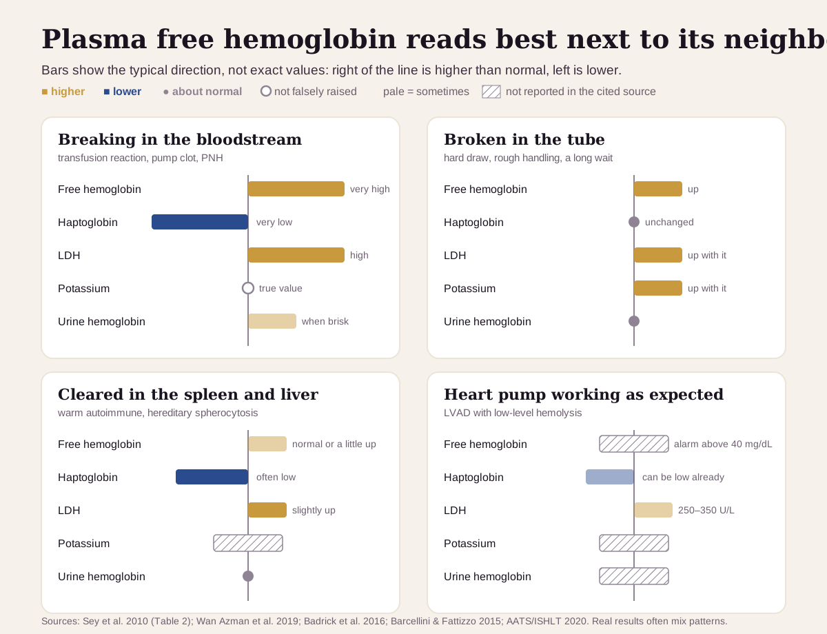
Plasma free hemoglobin on its own can't tell a broken tube from a broken vessel. Four other results can:24
| If you see… | It often points to… |
|---|---|
| High free hemoglobin, very low haptoglobin, high LDH, potassium not falsely raised | red cells breaking inside the vessels: a transfusion reaction, a pump or valve problem, PNH246 |
| High free hemoglobin with normal haptoglobin and high potassium | red cells broken in the tube; a careful redraw usually settles it4 |
| Low haptoglobin, LDH mildly up, free hemoglobin normal or barely up, no hemoglobin in the urine | red cells cleared in the spleen and liver, as in warm autoimmune hemolytic anemia28 |
| LDH around 250–350 U/L and a haptoglobin that may already be low in someone with an LVAD | the low-level hemolysis many pumps cause; after the first week, free hemoglobin above 40 mg/dL or an LDH over 2.5× the upper limit is the warning7 |
| Dark urine positive for blood with few or no red cells under the microscope | hemoglobin (or muscle myoglobin) in the urine; free hemoglobin, haptoglobin and LDH show whether red cells are breaking2 |
LDH is the slower, steadier partner: it comes out of many tissues, so it needs free hemoglobin or haptoglobin beside it to point at red cells.7 Our LDH guide walks through its other sources.
How fast plasma free hemoglobin changes
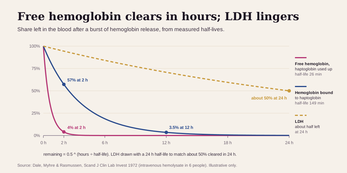
It clears within hours. In a classic experiment, loose hemoglobin injected into people whose haptoglobin was used up had a half-life of about 26 minutes. With haptoglobin on hand, the hemoglobin-haptoglobin pair had a half-life of about 149 minutes. LDH lingered: only about half of it had cleared after 24 hours.18
What that means in practice:
- Timing is everything. By these half-lives, a sample drawn a few hours after a short burst of hemolysis can already be back near normal. The study authors concluded that free hemoglobin is a poor gauge of how much hemolysis happened, and that LDH works better for that.18
- Hospitals track it on a schedule. Heart-pump guidelines suggest checking LDH and plasma free hemoglobin monthly, and ECMO studies check it in the first 24 hours.710
- Trends beat snapshots. A single high value on a pink sample needs a careful redraw. A value that climbs on repeat samples, with haptoglobin falling, is real.46
Going deeper: where loose hemoglobin goes
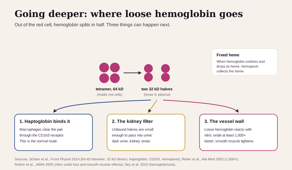
Inside red cells, hemoglobin is a four-part molecule (a tetramer) of about 64 kD. Loose in the plasma, it splits back and forth into 32 kD halves (dimers). Those halves are small enough to slip through the kidney's filter, unless haptoglobin grabs them first.11 That's why brisk hemolysis turns urine dark, and why kidney injury is linked to hemolysis.213
When hemoglobin loses its heme, a second protein, hemopexin, collects the heme.11 Loose hemoglobin also reacts with nitric oxide at least 1,000 times faster than hemoglobin inside red cells does.17 Losing nitric oxide tightens smooth muscle. In PNH, it's linked to spasms of the gut, narrowed blood vessels and clotting problems.9 One man with an LVAD had trouble swallowing and dark urine, a free hemoglobin of 125 mg/dL and a haptoglobin under 10; both symptoms went away once the hemolysis stopped.15
How labs see it: hemoglobin absorbs light at 415, 540 and 570 nm, so analyzers can measure a "hemolysis index" on each sample and flag broken ones.614 Very high bilirubin, cloudy or fatty plasma, and methemalbumin (which turns serum brown) can push some methods falsely high.36
Questions worth asking
- Was this sample drawn and handled for a plasma hemoglobin test, or could the tube be the cause?
- What did my haptoglobin, LDH and potassium show on the same draw?
- Should we repeat it with a careful draw before acting on it?
- If I have an LVAD or am on ECMO, what number would change the plan?
- Did I have a transfusion recently, and should a transfusion reaction be checked?
- Is my urine dark, and should it be tested for hemoglobin?
FAQ
What is a normal plasma free hemoglobin? Very low. Labcorp's range is under 5 mg/dL with a careful draw, and ARUP's is 0–9.7 mg/dL. Use the range printed on your report.35
Can a bad blood draw cause a high plasma free hemoglobin? Yes. Broken samples are the most common sample problem labs see. Red cells broken in the tube raise the result along with potassium and LDH, while haptoglobin stays normal. A careful redraw usually settles it.414
What level is dangerous? Heart-pump teams act above 40 mg/dL, ECMO teams use 50 mg/dL, and over 100 mg/dL counts as severe in children on ECMO. Dark urine with a high result needs same-day care.710122
What's the difference between plasma free hemoglobin and haptoglobin? Free hemoglobin is the loose hemoglobin itself, and it clears within hours.18 Haptoglobin is the protein that mops it up, and it stays low for 2 to 3 days after even 25 mL of blood breaks down.19 Read them together.
This guide is for education. It can't diagnose you or replace care from a clinician who knows your history.
Footnotes
-
National Library of Medicine. Hemoglobinuria test. MedlinePlus Medical Encyclopedia. Where old red cells are normally broken down, and when hemoglobin reaches the urine. MedlinePlus ↩ ↩2 ↩3
-
Sey MS, Manlucu J, Myers KA. Intravascular hemolysis secondary to aorto-atrial fistula presenting as red urine. J Gen Intern Med. 2010;25(12):1370–1374. Its Table 2 compares intravascular and extravascular hemolysis marker by marker. PMC ↩ ↩2 ↩3 ↩4 ↩5 ↩6 ↩7 ↩8 ↩9 ↩10 ↩11 ↩12 ↩13 ↩14 ↩15 ↩16 ↩17
-
Labcorp. Hemoglobin (Hb), Free, Plasma. Test 005595. A commercial laboratory's test page, cited for its reference range (under 5 mg/dL if conditions are optimal; 5–15 mg/dL with caution), uses, collection instructions, method and interferences. labcorp.com ↩ ↩2 ↩3 ↩4 ↩5 ↩6 ↩7 ↩8 ↩9 ↩10 ↩11 ↩12 ↩13 ↩14 ↩15 ↩16 ↩17 ↩18 ↩19
-
Wan Azman WN, Omar J, Koon TS, Tuan Ismail TS. Hemolyzed specimens: major challenge for identifying and rejecting specimens in clinical laboratories. Oman Med J. 2019;34(2):94–98. Causes of in-tube hemolysis and how labs tell it from hemolysis in the body. PMC ↩ ↩2 ↩3 ↩4 ↩5 ↩6 ↩7 ↩8 ↩9 ↩10 ↩11 ↩12 ↩13 ↩14 ↩15 ↩16
-
ARUP Laboratories. Hemoglobin, Plasma. Test 0020058. A commercial laboratory's test page (ARUP is a nonprofit academic reference lab), cited for its 0.0–9.7 mg/dL interval, heparin-plasma and 2-hour separation rules, and ordering notes. aruplab.com ↩ ↩2 ↩3 ↩4 ↩5 ↩6 ↩7 ↩8 ↩9 ↩10
-
Badrick T, Barden H, Callen S, et al. Consensus statement for the management and reporting of haemolysed specimens. Clin Biochem Rev. 2016;37(4):140–142. Australasian laboratory consensus, including when to suspect hemolysis in the body. PMC ↩ ↩2 ↩3 ↩4 ↩5 ↩6 ↩7 ↩8 ↩9
-
Kirklin JK, Pagani FD, Goldstein DJ, et al. American Association for Thoracic Surgery/International Society for Heart and Lung Transplantation guidelines on selected topics in mechanical circulatory support. J Heart Lung Transplant. 2020;39(3):187–219. The INTERMACS over-40 mg/dL definition, LDH thresholds and monitoring advice for heart pumps. ISHLT · PubMed ↩ ↩2 ↩3 ↩4 ↩5 ↩6 ↩7 ↩8 ↩9 ↩10 ↩11 ↩12 ↩13
-
Barcellini W, Fattizzo B. Clinical applications of hemolytic markers in the differential diagnosis and management of hemolytic anemia. Dis Markers. 2015;2015:635670. LDH slightly raised in extravascular hemolysis, 4–5 times the upper limit in PNH and prosthetic-valve hemolysis. PMC ↩ ↩2 ↩3 ↩4 ↩5
-
Rother RP, Bell L, Hillmen P, Gladwin MT. The clinical sequelae of intravascular hemolysis and extracellular plasma hemoglobin: a novel mechanism of human disease. JAMA. 2005;293(13):1653–1662. The review that tied loose hemoglobin to nitric oxide loss and its symptoms. PubMed ↩ ↩2 ↩3 ↩4 ↩5
-
Omar HR, Mirsaeidi M, Socias S, et al. Plasma free hemoglobin is an independent predictor of mortality among patients on extracorporeal membrane oxygenation support. PLoS One. 2015;10(4):e0124034. 154 adults on ECMO; uses the ELSO cutoff of 50 mg/dL. PMC ↩ ↩2 ↩3 ↩4 ↩5 ↩6 ↩7 ↩8 ↩9
-
Schaer DJ, Vinchi F, Ingoglia G, Tolosano E, Buehler PW. Haptoglobin, hemopexin, and related defense pathways: basic science, clinical perspectives, and drug development. Front Physiol. 2014;5:415. PMC ↩ ↩2 ↩3 ↩4 ↩5
-
Alexander PMA, Di Nardo M, Combes A, et al. Definitions of adverse events associated with extracorporeal membrane oxygenation in children: results of an international Delphi process from the ECMO-CENTRAL ARC. Lancet Child Adolesc Health. 2024;8(10):773–780. Moderate hemolysis 50–100 mg/dL, severe over 100 mg/dL (82% agreement). PMC · ECMO-CENTRAL definitions ↩ ↩2 ↩3 ↩4
-
Vermeulen Windsant IC, de Wit NC, Sertorio JT, et al. Hemolysis during cardiac surgery is associated with increased intravascular nitric oxide consumption and perioperative kidney and intestinal tissue damage. Front Physiol. 2014;5:340. 67 adults having heart surgery. PMC ↩ ↩2 ↩3
-
Lippi G, Blanckaert N, Bonini P, et al. Haemolysis: an overview of the leading cause of unsuitable specimens in clinical laboratories. Clin Chem Lab Med. 2008;46(6):764–772. PubMed ↩ ↩2 ↩3
-
Wuschek A, Iqbal S, Estep J, Quigley E, Richards D. Left ventricular assist device hemolysis leading to dysphagia. World J Gastroenterol. 2015;21(18):5735–5738. A single case report. PMC ↩ ↩2
-
National Library of Medicine. Hemolytic transfusion reaction. MedlinePlus Medical Encyclopedia. Symptoms and timing. MedlinePlus ↩ ↩2
-
Reiter CD, Wang X, Tanus-Santos JE, et al. Cell-free hemoglobin limits nitric oxide bioavailability in sickle-cell disease. Nat Med. 2002;8(12):1383–1389. PubMed ↩ ↩2 ↩3
-
Dale J, Myhre E, Rasmussen K. Elimination of hemoglobin and lactate dehydrogenase from plasma in normals and patients with intravascular hemolysis. Scand J Clin Lab Invest. 1972;29(4):389–395. The half-lives of free hemoglobin (26 min) and the hemoglobin-haptoglobin pair (149 min). PubMed ↩ ↩2 ↩3 ↩4
-
Labcorp. Haptoglobin. Test 001628. A commercial laboratory's test page: "Haptoglobin is decreased for two to three days after only 25 mL of blood is lysed." labcorp.com ↩
Educational content, not medical advice. Libby is a personal record tool, not a medical service — it doesn't diagnose, treat, or prescribe. Reference ranges vary by lab and by person. Talk to a qualified healthcare professional about your results.
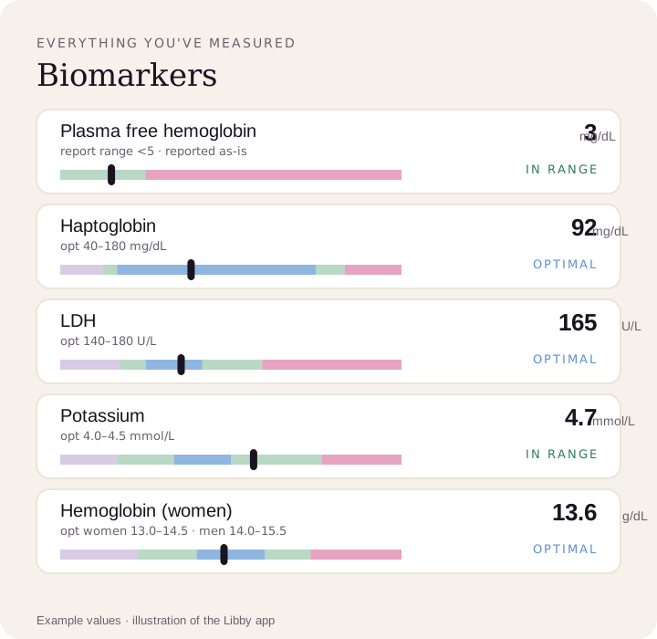
See every result against your lab's range.
Upload a lab PDF or a photo of your report. Libby shows your plasma free hemoglobin as your lab reports it, next to your haptoglobin, LDH and potassium, and tracks each new result.
View your plasma free hemoglobin ›