Hemoglobin A percentage: normal range and what low means
Hemoglobin A is the main adult hemoglobin, usually 95–98% of the total. How the test works, and what a lower share means, from carrier traits to transfusion.
Hemoglobin A (HbA) is the main hemoglobin in adult red blood cells. On a hemoglobin electrophoresis report it's usually 95–98% of the total.1 The test sorts your hemoglobin into its types and gives each one's share. Because the shares add up to 100%, HbA is what's left over: when another type rises, such as sickle hemoglobin (HbS), HbA2 or fetal hemoglobin (HbF), the HbA percentage falls.
The short version
- Typical adult: HbA 95–98%, HbA2 2–3%, HbF under 2%, and no HbS, HbC or HbE.12 Labcorp's range for HbA from age 2 is 96.4–98.8%.3
- A few points low usually means HbA2 has risen above about 3.5%, the pattern of beta-thalassemia trait, a carrier state.45
- About 60% HbA with about 40% HbS is sickle cell trait: one copy of the sickle gene, and most people with it have no symptoms.16 0% HbA with mostly HbS points to sickle cell disease.1
- A blood transfusion in the past 12 weeks adds donor HbA and can make the result misleading. Tell the lab if you've had one.2
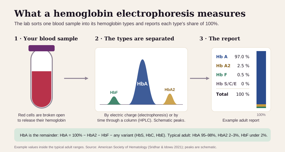
What hemoglobin A is
Hemoglobin is the oxygen-carrying protein that fills your red cells. Each molecule is built from four protein chains. HbA has two alpha and two beta chains. HbA2 swaps the beta chains for delta chains, and HbF swaps them for gamma chains.1
Before birth, HbF does most of the work. Shortly after birth your body switches from making gamma chains to making beta chains, so HbA takes over.7 By age 1 to 2, HbA has usually replaced most of the HbF.2
HbA1c is a different test. It measures how much of your hemoglobin has glucose stuck to it, a gauge of your blood sugar over the past two to three months.8 That's covered in our HbA1c guide.
You'll usually see HbA on a hemoglobin electrophoresis or hemoglobinopathy evaluation. Common reasons for the test:
- Newborn screening, which checks babies for sickle cell disease and other hemoglobin disorders shortly after birth.2
- Planning a pregnancy. About 1 in 66 people in the U.S. carry a hemoglobin trait, so the American College of Obstetricians and Gynecologists recommends offering the test to everyone planning a pregnancy or at the first prenatal visit.9
- Symptoms of anemia or sickle cell disease, or a family history of a hemoglobin disorder.2 Labcorp also asks for your MCV (average red-cell size) and transfusion history with the order.3
How the lab measures it. The lab breaks open red cells and separates the hemoglobin types by their electric charge (electrophoresis) or by how long each takes to pass through a column (HPLC, high-performance liquid chromatography). Standard practice is two methods: one to screen and one to confirm anything unusual.1 Labcorp starts with capillary electrophoresis; ARUP starts with HPLC.310 You don't need to fast.3
What's a normal hemoglobin A?
In adults, the usual pattern is HbA 95–98%, HbA2 2–3% and HbF under 2%, according to the American Society of Hematology's teaching table.1 MedlinePlus gives HbA as more than 95%, and HbS, HbC and HbE as absent.2
Each lab sets its own range, because the machines differ:
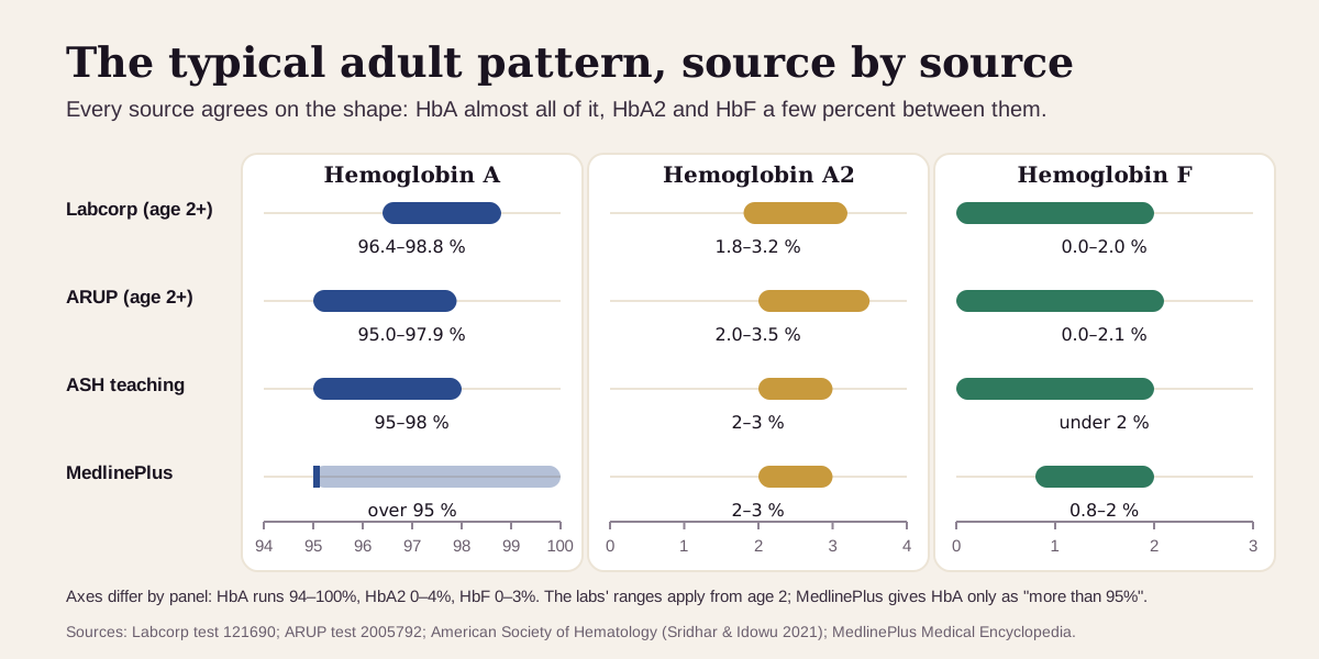
- Labcorp uses 96.4–98.8% for HbA from age 2, and 94.6–98.5% from 7 months to 1 year.3
- ARUP uses 95.0–97.9% from age 2.10
- In a 2023 study of 321 people without a hemoglobin disorder, the middle 95% of HbA results ran 97.0–98.0% by HPLC and 96.6–98.4% by capillary electrophoresis.4
Men and women share the same range. None of these sources split HbA by sex. Age matters a lot in the first two years, though: in a baby's first month, ARUP's range for HbA is 7.6–54.8%, because HbF still makes up most of the total.10 Labcorp sets no HbA range before 7 months.3
"Optimal" hemoglobin A: there's no target to chase
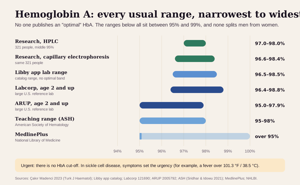
Health optimizers set "optimal" bands for many markers. HbA has none, and for a good reason: your share of HbA is set by the globin genes you inherited, so the useful question is whether your pattern is the usual adult one, and if not, what took HbA's place. Here is every published "usual" range, from narrowest to widest:
| Range | Hemoglobin A | Who uses it |
|---|---|---|
| Strictest (longevity platforms) | none published | Function Health and Superpower list HbA1c and total hemoglobin, but no HbA share11 |
| Research, HPLC | 97.0–98.0% | Middle 95% of 321 people without a hemoglobin disorder4 |
| Research, capillary electrophoresis | 96.6–98.4% | The same 321 people4 |
| Libby app lab range | 96.5–98.5% | The lab range in the Libby app's marker catalog, with no separate optimal band |
| Labcorp, age 2 and up | 96.4–98.8% | A large U.S. reference lab3 |
| ARUP, age 2 and up | 95.0–97.9% | A large U.S. reference lab10 |
| Teaching range | 95–98% | American Society of Hematology primer1 |
| Consumer reference | over 95% | MedlinePlus2 |
| Urgent | no HbA cut-off | In sickle cell disease, symptoms set the urgency, e.g. a fever over 101.3 °F (38.5 °C)6 |
What the ladder shows: every range sits between about 95% and 99%. The two research ranges come from the same people measured on two machines, and their medians differ by just 0.1 point (97.4% vs 97.5%).4 So a few tenths up or down inside the range mostly reflects the lab's method. Compare results from the same lab.
Keep "optimal" in perspective: a higher HbA is no healthier than a normal one. A result a few points low, or far lower, is information about which hemoglobin types you carry, and it's worth pairing with your blood count and family history.
What a lower hemoglobin A usually means
Start with what took HbA's place. HbA is the remainder, so the other fractions on the same report tell you why it's low.
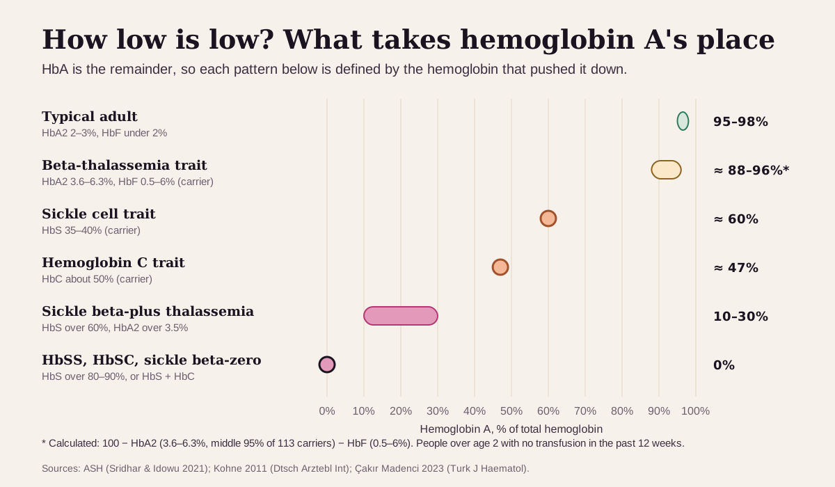
A few points low: more HbA2 or HbF
- Beta-thalassemia trait. This carrier state means one beta-globin gene makes less beta chain. HbA2 rises above about 3.5% (a median of 5% in 113 people with the trait in one study), and red cells are small, with an MCV of 55–75 fL. HbA lands a few points low, roughly 88–96%. Mild anemia is common.45 Our HbA2 guide covers the cut-offs.
- More fetal hemoglobin. Some people inherit hereditary persistence of fetal hemoglobin, which keeps HbF high into adulthood. People with sickle cell disease who also carry it have had few symptoms.7 The sickle cell medicine hydroxyurea raises HbF on purpose: in the multicenter hydroxyurea study, HbF reached 18.1% after 2 years in the quarter of patients who responded best.12 More in our HbF guide.
- A baby or toddler. HbA is still climbing toward adult levels until about age 2.10
About half to two-thirds: a carrier trait
- Sickle cell trait. HbA and HbS usually split about 60:40.1 It's a carrier state: one copy of the sickle gene. More than 2 million people in the U.S. have it, most have no symptoms and lead normal lives, and the trait never turns into sickle cell disease.6 Problems are rare and tend to come with extreme conditions: intense exercise, high altitude, dehydration, or extreme heat or cold.6 See our HbS guide and the sickle cell screen guide.
- Hemoglobin C trait. HbC about 50% and HbA about 47%, with no apparent illness.5
- Hemoglobin E trait. HbE takes 25–35%.5 It usually causes mild anemia or none.2
Very low or zero: sickle cell disease and related conditions
If your body makes no normal beta chains, you have no HbA at all. That's the pattern in sickle cell anemia (HbSS, with HbS over 90%), hemoglobin SC disease (HbS and HbC about 50% each) and sickle beta-zero thalassemia.1 In sickle beta-plus thalassemia, some normal beta chain gets through and HbA runs 10–30%.1 Sickle cell disease affects nearly 100,000 people in the U.S.13 Telling these conditions apart takes the full pattern, a blood count, and sometimes gene testing.110
Higher than your own body makes: a recent transfusion
Donor red cells carry HbA. So in someone with sickle cell anemia, any HbA on the report came from a transfusion. In one man with sickle cell anemia, a red-cell exchange raised HbA from 63.0% to 71.5% and cut HbS from 33.5% to 11.0%.14 Long-term transfusion programs aim to keep HbS under 30% before the next transfusion.13 For anyone, a transfusion in the past 12 weeks can make the result falsely normal or abnormal.2
Get care now if you have sickle cell disease and a fever over 101.3 °F (38.5 °C), severe pain, chest pain with cough or shortness of breath, extreme tiredness with dizziness or an irregular heartbeat, or an erection lasting 4 hours or more. Call 911 for sudden weakness or numbness on one side, confusion, or trouble speaking, seeing or walking.6
Hemoglobin A reads best next to its neighbors
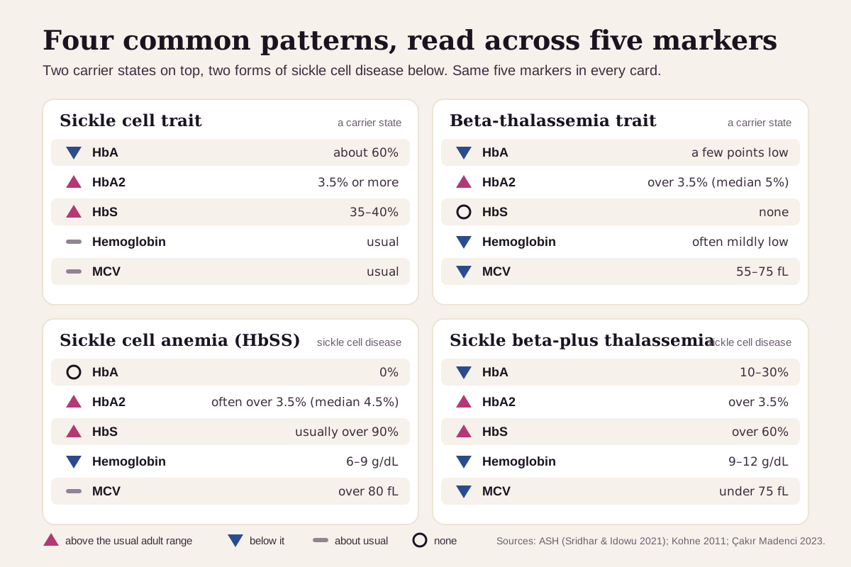
HbA makes sense only next to the other fractions and your blood count. Four common patterns:
| If you see… | It often points to… |
|---|---|
| HbA a few points low, HbA2 above 3.5%, low MCV, no HbS | beta-thalassemia trait, a carrier state |
| HbA about 60%, HbS about 40%, normal blood count | sickle cell trait, a carrier state |
| HbA 0%, HbS over 90%, hemoglobin 6–9 g/dL, MCV over 80 fL | sickle cell anemia (HbSS) |
| HbA 10–30%, HbS over 60%, HbA2 above 3.5%, MCV under 75 fL | sickle beta-plus thalassemia |
| HbA 0%, HbS and HbC about 50% each | hemoglobin SC disease |
| HbA about 47%, HbC about 50% | hemoglobin C trait |
| HbS with high HbF and a high MCV | often hydroxyurea treatment |
| HbA present in someone known to have HbSS | a recent transfusion |
| Normal HbA, HbA2 and HbF, but small red cells | possible alpha-thalassemia trait, which shows a normal pattern on this test |
Low iron also makes red cells small, so clinicians rule out iron deficiency before reading a low MCV as a thalassemia sign.1
How fast hemoglobin A changes
In an adult, the pattern holds steady. It comes from the globin genes you inherited,7 and from age 2 the usual range is narrow, such as Labcorp's 96.4–98.8%.3 Three things move it:
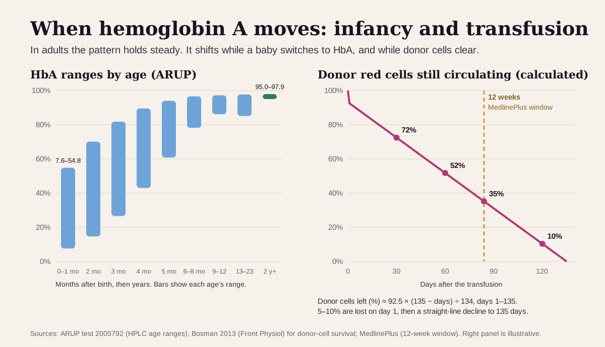
- Infancy. HbA climbs from a minority of the total at birth to the adult range by age 2.10 ARUP calls results most reliable from age 1.10
- A transfusion. Red cells live about 120 days on average.16 Donor cells last up to 135 days, with a straight-line decline after the first day.15 By that math, about a third of the donor cells are still there at 12 weeks, which is why MedlinePlus flags transfusions in that window.2
- Hydroxyurea. HbF rises over months of treatment, and the multicenter study tracked the response over 2 years.12
Going deeper: one protein, several versions
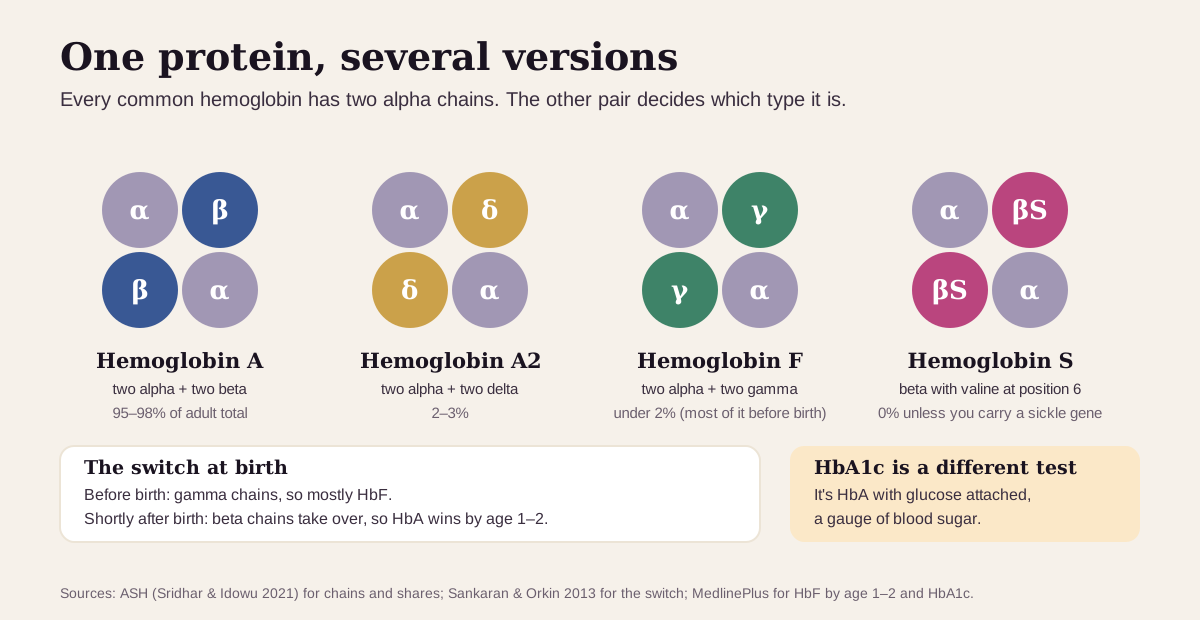
All the common hemoglobins share the same two alpha chains. What differs is the other pair: beta in HbA, delta in HbA2 and gamma in HbF.1 Before birth, the gamma genes are switched on. Shortly after birth, a genetic switch turns gamma down and beta up.7
HbS is HbA with one change. At position 6 of the beta chain, the amino acid valine replaces glutamic acid.1 In sickle cell trait the split is closer to 60:40 than 50:50, because alpha chains pair more readily with normal beta chains.1
Why two methods? Electrophoresis sorts hemoglobin by charge. HPLC sorts it by how long each type takes to come off a column, and it measures HbA2 and HbF precisely. Some variants land in the same spot on one method (HPLC can blur HbE with HbA2, for example), so labs confirm unusual results with a second one.14
HbA1c is HbA with glucose attached. The more sugar in your blood over the past few months, the more of it you'll have.8
Questions worth asking
- Which fraction took HbA's place: HbA2, HbF, HbS or another variant?
- Could a transfusion in the past 3 months affect this result?
- Was a second method or a gene test used to confirm the pattern?
- How do my hemoglobin, MCV and iron results fit with it?
- If this is a carrier trait, should my partner be tested before a pregnancy?
- Should my parents, siblings or children be tested too?
FAQ
What is a normal hemoglobin A percentage? In adults, about 95–98% of total hemoglobin.1 Labcorp uses 96.4–98.8% from age 2.3
What does a low hemoglobin A mean? Another hemoglobin type has taken a bigger share. A few points low usually means a higher HbA2 (beta-thalassemia trait), about 60% usually means sickle cell trait, and 0% points to sickle cell disease.15
Is hemoglobin A the same as HbA1c? No. HbA is a share of your hemoglobin types. HbA1c measures how much of your hemoglobin has glucose attached, which tracks blood sugar.8
Can a blood transfusion change my hemoglobin A? Yes. Donor red cells carry HbA and last up to about 135 days, so tell the lab if you've had a transfusion in the past 12 weeks.215
This guide is for education. It can't diagnose you or replace care from a clinician who knows your history.
Footnotes
-
Sridhar A, Idowu M. Hemoglobin Electrophoresis in Sickle Cell Disease: A Primer for the Clinician. American Society of Hematology, Hematopoiesis; November 16, 2021. The source for the adult pattern (HbA 95–98%, HbA2 2–3%, HbF under 2%), the chain make-up, the two-method standard, the 60:40 trait split, and the classic sickle cell disease patterns (a table from the ASH Self-Assessment Program). hematology.org ↩ ↩2 ↩3 ↩4 ↩5 ↩6 ↩7 ↩8 ↩9 ↩10 ↩11 ↩12 ↩13 ↩14 ↩15 ↩16 ↩17 ↩18 ↩19 ↩20 ↩21 ↩22 ↩23 ↩24 ↩25 ↩26
-
National Library of Medicine. Hemoglobin Electrophoresis. MedlinePlus lab test page and Medical Encyclopedia. Adult values (HbA more than 95%, HbA2 2–3%, HbF 0.8–2%), newborn screening, HbE, and the 12-week transfusion window. Lab test page · Encyclopedia ↩ ↩2 ↩3 ↩4 ↩5 ↩6 ↩7 ↩8 ↩9 ↩10 ↩11 ↩12 ↩13
-
Labcorp. Hemoglobinopathy Fractionation Cascade, test 121690. A commercial reference laboratory's published test page, opened September 23, 2026: HbA 96.4–98.8% from age 2 and 94.6–98.5% from 7 months to 1 year, capillary electrophoresis with reflex HPLC, no fasting. labcorp.com ↩ ↩2 ↩3 ↩4 ↩5 ↩6 ↩7 ↩8 ↩9 ↩10 ↩11
-
Çakır Madenci Ö, Hürmeydan Ö, Orçun A, Erdoğmuş F. Comparison of Capillary Zone Electrophoresis with High-pressure Liquid Chromatography in the Evaluation of Hemoglobinopathies. Turk J Haematol. 2023;40(4):258–265. 321 people without a hemoglobin disorder measured on both methods, plus 113 with beta-thalassemia trait. PMC ↩ ↩2 ↩3 ↩4 ↩5 ↩6 ↩7 ↩8 ↩9 ↩10 ↩11
-
Kohne E. Hemoglobinopathies: clinical manifestations, diagnosis, and treatment. Dtsch Arztebl Int. 2011;108(31–32):532–540. Tables of typical hemoglobin patterns and blood counts for thalassemia traits, sickle cell trait, and hemoglobin C and E traits. PMC ↩ ↩2 ↩3 ↩4 ↩5 ↩6 ↩7 ↩8
-
National Heart, Lung, and Blood Institute. Sickle Cell Trait (updated August 22, 2024) and Sickle Cell Disease: Symptoms (updated August 20, 2024). Trait facts and the emergency warning signs, including a fever over 101.3 °F (38.5 °C). Sickle cell trait · Symptoms ↩ ↩2 ↩3 ↩4 ↩5
-
Sankaran VG, Orkin SH. The switch from fetal to adult hemoglobin. Cold Spring Harb Perspect Med. 2013;3(1):a011643. The gamma-to-beta switch around birth and hereditary persistence of fetal hemoglobin. PMC ↩ ↩2 ↩3 ↩4 ↩5
-
National Library of Medicine. Hemoglobin A1C (HbA1c) Test. MedlinePlus. Glucose sticks to hemoglobin; the test measures the share of glucose-coated hemoglobin. MedlinePlus ↩ ↩2 ↩3 ↩4
-
American College of Obstetricians and Gynecologists. Hemoglobinopathies in Pregnancy. Practice Advisory; August 2022. About 1 in 66 people in the U.S. have a hemoglobin trait; testing offered to everyone planning pregnancy or at the first prenatal visit. acog.org ↩
-
ARUP Laboratories. Hemoglobin Evaluation Reflexive Cascade, test 2005792. A reference laboratory's commercial test directory, opened September 23, 2026: HPLC-first testing and age-specific HbA ranges from 7.6–54.8% (first month) to 95.0–97.9% (age 2 and up). aruplab.com ↩ ↩2 ↩3 ↩4 ↩5 ↩6 ↩7 ↩8 ↩9 ↩10 ↩11
-
Function Health, "What we test," and Superpower, "Biomarkers." Commercial testing companies' public biomarker lists, opened September 23, 2026. Both list HbA1c and hemoglobin, and neither lists a hemoglobin A, A2 or F fraction or target. functionhealth.com · superpower.com ↩
-
Steinberg MH, Lu ZH, Barton FB, Terrin ML, Charache S, Dover GJ. Fetal hemoglobin in sickle cell anemia: determinants of response to hydroxyurea. Blood. 1997;89(3):1078–1088. 150 patients in the Multicenter Study of Hydroxyurea; HbF reached 18.1% and 8.8% at 2 years in the top two response quarters. PubMed ↩ ↩2 ↩3
-
Yawn BP, Buchanan GR, Afenyi-Annan AN, et al. Management of sickle cell disease: summary of the 2014 evidence-based report by expert panel members. JAMA. 2014;312(10):1033–1048. The NHLBI expert panel: nearly 100,000 people with sickle cell disease in the U.S., and the HbS-under-30% transfusion target. PubMed ↩ ↩2
-
Swedan N, Nicol K, Moder P, Kahwash S. An "acquired" hemoglobin J variant in a sickle cell disease patient. Ther Clin Risk Manag. 2008;4(3):649–652. Before-and-after hemoglobin fractions for one red-cell exchange. PMC ↩ ↩2
-
Bosman GJ. Survival of red blood cells after transfusion: processes and consequences. Front Physiol. 2013;4:376. Donor cells survive up to 135 days; 5–10% disappear in the first 24 hours, then decline in a straight line. PMC ↩ ↩2 ↩3
-
Thiagarajan P, Parker CJ, Prchal JT. How Do Red Blood Cells Die? Front Physiol. 2021;12:655393. Red cells live about 120 days on average. PMC ↩
Educational content, not medical advice. Libby is a personal record tool, not a medical service — it doesn't diagnose, treat, or prescribe. Reference ranges vary by lab and by person. Talk to a qualified healthcare professional about your results.
See every result against your lab's range.
Upload a lab PDF or a photo of your report. Libby puts every marker on your lab's range, adds the optimal range where one exists, and tracks your trends over time.
View your hemoglobin A ›