Fetal hemoglobin (HbF): normal levels and what high means
Hemoglobin F is the hemoglobin you made before birth. Normal levels by age, why it runs high in HPFH and thalassemia, and why more helps in sickle cell.
Hemoglobin F (HbF), or fetal hemoglobin, is the hemoglobin your body made before you were born. In the months after birth your marrow switches to adult hemoglobin (HbA), so from age 2 HbF is usually 2% or less of the total.12 A higher level in an older child or adult often points to something inherited: a thalassemia trait, sickle cell disease, or a harmless trait called hereditary persistence of fetal hemoglobin (HPFH).3 In sickle cell disease, more HbF is protective.4
The short version
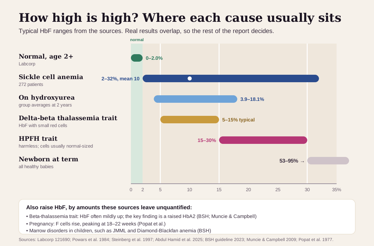
- Normal (age 2 and up): 0.0–2.0% at Labcorp. MedlinePlus lists 0.8–2% for adults.25 Babies are different: at birth HbF is 53–95% of all hemoglobin, and it falls over the first 6–12 months.3
- Raised in an adult often means an inherited pattern. Delta-beta thalassemia trait typically runs about 5–15% with small red cells, and HPFH about 15–30%, usually with normal-sized cells and no illness.6 In beta-thalassemia trait, HbF can be mildly raised, but the telling number is HbA2.3
- In sickle cell disease, higher is better. In 272 people with sickle cell anemia, HbF above about 10% went with fewer strokes and organ complications, and above 20% with fewer pain crises.4 In the main trial of the medicine hydroxyurea, about half of the adults taking it kept a higher HbF over 2 years.7
- A separate test: the Kleihauer-Betke test in pregnancy uses HbF to count a baby's red cells in the mother's blood. It answers a different question from your own HbF result.8
What hemoglobin F is
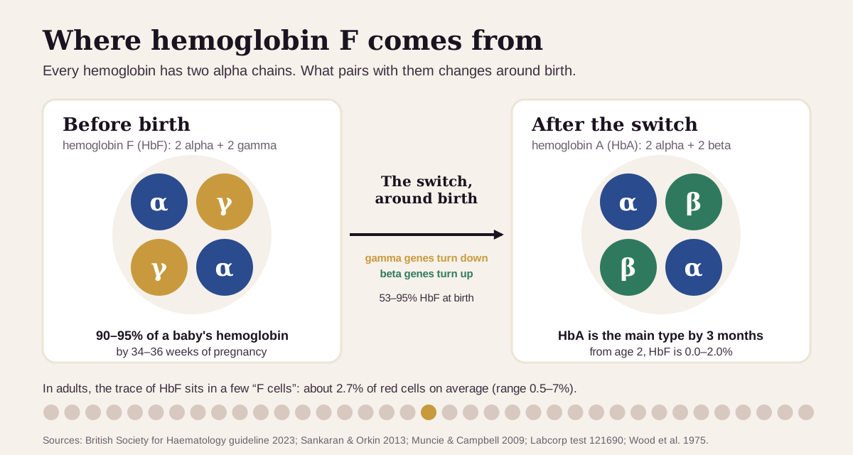
Hemoglobin is the oxygen-carrying protein inside red cells. Each molecule is built from four chains: two alpha chains and two partners. HbF pairs the alphas with two gamma chains. Adult HbA pairs them with two beta chains, and the minor adult type, HbA2, with two delta chains.9
Before birth, gamma chains dominate: HbF is 90–95% of a baby's hemoglobin by 34–36 weeks of pregnancy.3 Around birth a genetic switch turns the gamma-globin genes down and the beta-globin genes up.1 By 3 months, HbA is the main hemoglobin.3 (More on the switch at the end.)
In adults, the trace of HbF that remains sits in a minority of red cells called F cells. In healthy adults they average 2.7% of red cells (range 0.5–7%), and the more F cells you have, the higher your HbF.10
HbF appears as one line on a hemoglobin electrophoresis report (Labcorp calls it hemoglobinopathy fractionation), next to HbA, HbA2 and any variants. Labs use it to look for thalassemias and hemoglobin variants,2 for example when a blood count shows small red cells with no clear cause.3 Our hemoglobin A guide walks through the whole report.
What's a normal hemoglobin F?
Age matters more than anything else. Here's how the published ranges line up:352
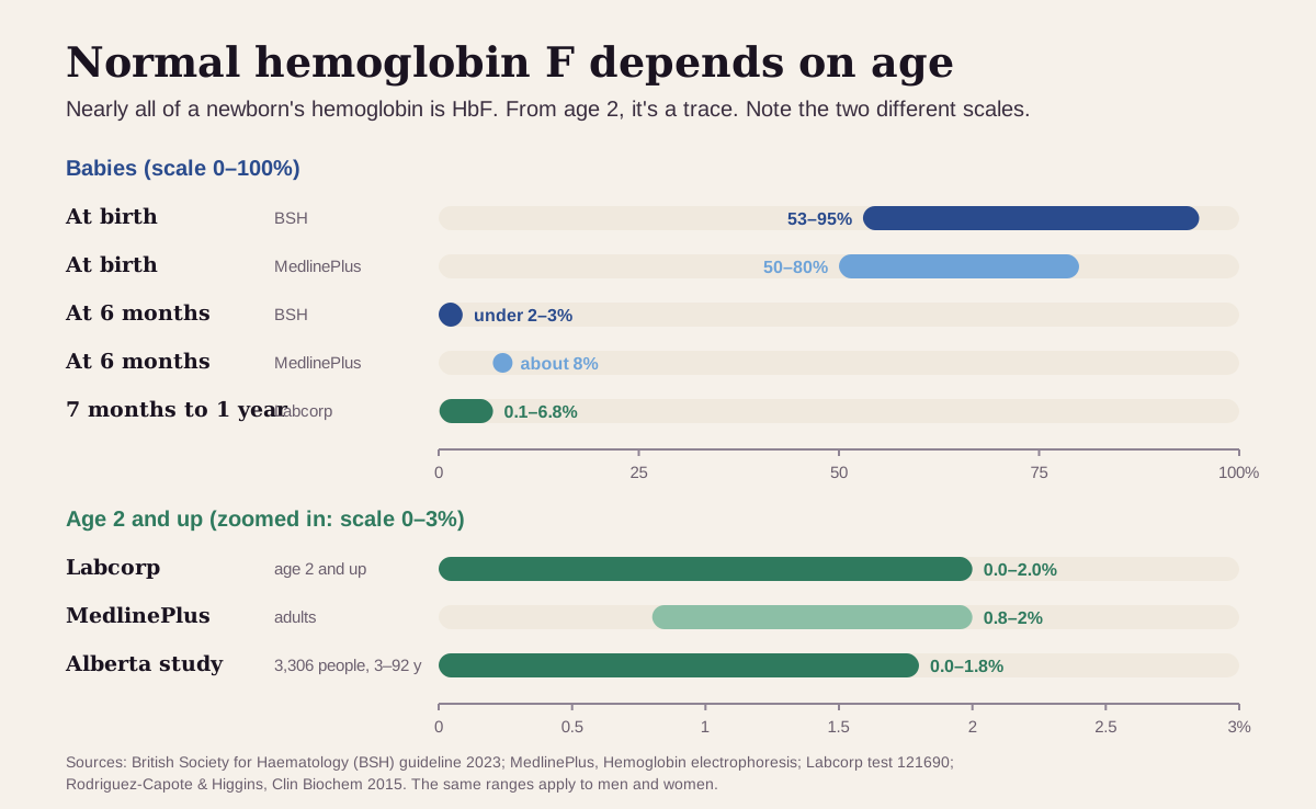
- At birth: the British Society for Haematology (BSH) puts HbF at 53–95% of all hemoglobin at term, with a typical newborn sample around 75%.3 MedlinePlus lists 50–80%.5
- The first year: HbF falls fast. Labcorp's range for 7 months to 1 year is 0.1–6.8%, and it prints no range under 7 months.2
- Age 2 and up: Labcorp uses 0.0–2.0%.2 A Canadian lab study of 3,306 people aged 3 to 92 with normal hemoglobin studies found 0.0–1.8%.11 A result of 0, or "none detected," is normal.
These sources give one range for men and women. Some babies start higher: those born small for their gestational age, those who have had long-term low oxygen, and babies with trisomy 13.3 Babies of mothers with diabetes make the switch later.1
"Optimal" hemoglobin F: what the ranges say
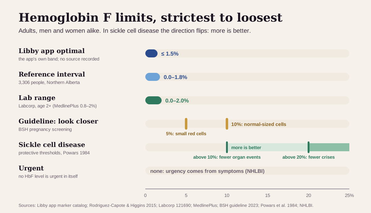
For most markers, health optimizers aim tighter than the lab. For HbF, the tightest band is the Libby app's own, and the one setting where a number is a goal rather than a limit is sickle cell disease, where more is better. Here is the adult ladder, strictest first:
| Target | Adults (men and women) | Who uses it |
|---|---|---|
| Libby app optimal | 1.5% or less | The optimal band shown in the Libby app. The app's catalog records no published source for this band, and it uses the same band at every age |
| Research reference interval | 0.0–1.8% | 3,306 people aged 3–92 with normal hemoglobin studies in Northern Alberta11 |
| Lab range | 0.0–2.0% (Labcorp); 0.8–2% (MedlinePlus) | Labcorp, age 2 and up;2 MedlinePlus's adult range5 |
| Guideline "look closer" points | 5% when MCH is under 27 pg (small, pale cells); 10% when MCH is normal | The BSH's pregnancy-screening triggers for delta-beta thalassemia and HPFH3 |
| Sickle cell disease: protective levels | above about 10% and 20% | Powars et al.: fewer major organ events above 10%, fewer recurring crises above 20%4 |
| Urgent | none | No HbF level is urgent in itself. In sickle cell disease, urgency comes from symptoms, such as a fever over 101.3°F (38.5°C)12 |
Why more HbF helps in sickle cell disease:
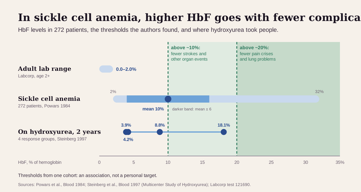
- Powars and colleagues followed 272 people with sickle cell anemia for an average of 11 years. Their HbF ranged from 2% to 32% (mean 10%). The authors found thresholds: about 10% for major organ events such as stroke or bone damage (aseptic necrosis), and about 20% for recurring events such as pain crises and lung problems.4
- In a study of 3,764 patients, a low HbF was one of the factors linked to early death in adults with sickle cell anemia, and a high HbF predicted better survival.13 An earlier analysis of 18,356 patient-years linked high pain rates to low HbF.14
- In the randomized Multicenter Study of Hydroxyurea, 299 adults with frequent crises had a median of 2.5 crises a year on hydroxyurea versus 4.5 on placebo. Fewer had acute chest syndrome (25 vs 51) or needed transfusions (48 vs 73).15 After 2 years, HbF in the four hydroxyurea response groups averaged 18.1%, 8.8%, 4.2% and 3.9%, so the rise varies a lot from person to person.7
Keep "optimal" in perspective. The app's 1.5% band describes a typical adult, and it's one band for everyone. So a baby's normal HbF and a hoped-for rise in someone with sickle cell disease both show as HIGH in the app. For a raised HbF from HPFH, the number is simply a trait: people who carry HPFH, even two copies, are healthy.63 What a raised HbF means depends on the rest of your report, which is what the next sections cover.
What a high hemoglobin F usually means
How high it is, and what the rest of your report shows, are the clues.3
The usual suspects
- Beta-thalassemia trait. A common carrier state. HbF is often mildly raised, but the finding that clinches it is a raised HbA2 with small red cells, usually an MCV under 75 fL.39 Our hemoglobin A2 guide covers the cutoffs. In one Alberta lab's year of testing, beta-thalassemia trait turned up 423 times and delta-beta thalassemia trait 10 times.11
- Sickle cell disease. HbF varies widely, from 2% to 32% in one large study, with an average of 10%.4 It rises further in many people who take hydroxyurea.7 See our guides to hemoglobin S and the sickle cell screen.
- Pregnancy. F cells rise during a normal pregnancy. In a study of 11 pregnancies, they rose in every one and peaked at 18–22 weeks.16 The BSH's pregnancy screening uses HbF of 5% or 10% (depending on the MCH) as its points to look closer.3
Less common: inherited high-HbF traits
- Delta-beta (δβ) thalassemia trait. A large deletion in the beta-globin gene cluster on one chromosome, with gamma chains partly filling the gap. HbF is typically about 5–15%, HbA2 is normal (under 3.0%), and red cells are small and pale. In one Malaysian series of 534 carriers, the delta-beta groups averaged about 14–21%.6 Alone it's a carrier state. Paired with a beta-thalassemia gene from the other parent, it can cause thalassemia intermedia or major,6 which is why the BSH asks for the baby's biological father to be tested when it's found in pregnancy.3
- HPFH (hereditary persistence of fetal hemoglobin). The gamma genes stay switched on in adult life: HbF is about 15–30%, red-cell indices are usually normal, and there's no illness. People with two copies are healthy too.6 The BSH calls it a benign condition.3 Inherited together with one sickle cell gene, HPFH makes for a far milder picture: the rare people with both were largely symptom-free.1
Rare but important
- Marrow disorders in children. Beyond infancy, the BSH lists juvenile myelomonocytic leukemia (JMML) and Diamond-Blackfan anemia among the conditions where a raised HbF should be measured.3
- Beta-thalassemia major. Babies are protected at birth by their HbF and develop symptoms as it falls, by about 6 months of age.91
Get care right away if you have sickle cell disease and develop a fever over 101.3°F (38.5°C); chest pain, cough or shortness of breath; severe pain; sudden weakness or numbness on one side, confusion, or trouble speaking, seeing or walking (call 911); or an erection lasting 4 hours or more. These are emergencies whatever your HbF.12
Hemoglobin F reads best next to its neighbors
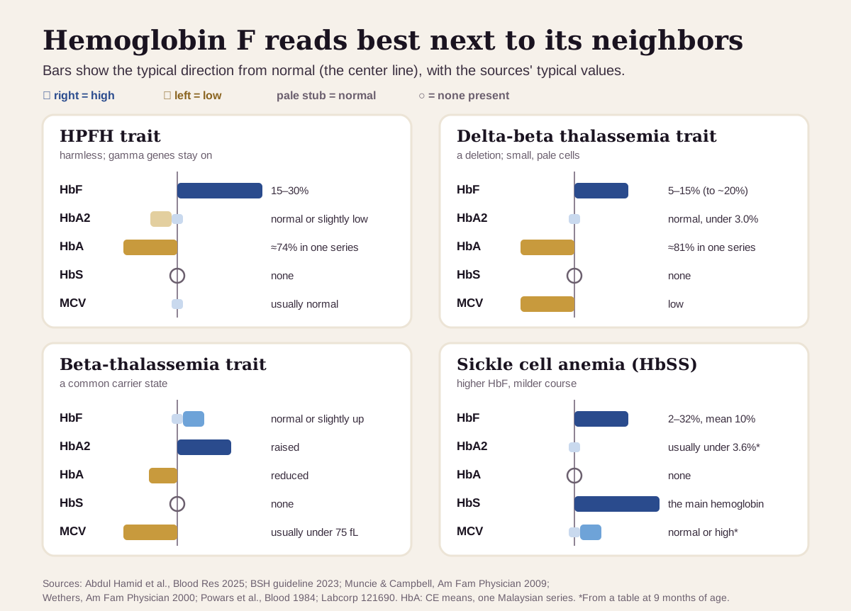
A raised HbF by itself leaves the cause open. The pattern across HbA2, HbA, any HbS and your red-cell size (MCV) does most of the sorting:36
| If you see… | It often points to… |
|---|---|
| HbF about 15–30%, HbA2 normal or slightly low, MCV usually normal | HPFH trait, a harmless inherited trait |
| HbF typically 5–15% (to about 20% in one large series), HbA2 normal (under 3.0%), small, pale red cells | delta-beta thalassemia trait |
| HbA2 raised, HbF normal or slightly up, MCV low (usually under 75 fL) | beta-thalassemia trait (see hemoglobin A2) |
| HbS the main hemoglobin, no HbA, HbF raised, MCV normal or high | sickle cell anemia (HbSS); a higher HbF tends to mean a milder course18 |
The sickle cell trait pattern, with more HbA than HbS, is covered in the sickle cell screen guide.17 To tell HPFH from delta-beta thalassemia, UK labs now use DNA testing in place of the older Kleihauer method, and flow cytometry can count F cells.3 Anemia and red-cell size have their own guides: hemoglobin and hematocrit, MCV and the reticulocyte count.
A separate test: fetal cells in a mother's blood
The Kleihauer-Betke test also relies on HbF, but it measures something else: how much of a baby's blood has crossed into the mother's circulation. Its main job is to help protect an Rh-negative mother, by sizing the Rh immune globulin that keeps her from making antibodies against an Rh-positive baby's red cells.8
It works because fetal red cells are packed with HbF, which survives an acid wash that strips adult hemoglobin out of the mother's own cells. The test is cheap but imprecise, and it can be thrown off when the mother has many F cells of her own. Flow cytometry with an anti-HbF antibody is the main alternative.8 Healthy adults carry some F cells (0.5–7% of red cells), and pregnancy raises them.1016 So a Kleihauer-Betke result in your records is about the pregnancy, and your hemoglobin electrophoresis HbF is about you.
How fast hemoglobin F changes
In babies, fast. In adults, slowly, around a set point that's mostly inherited.
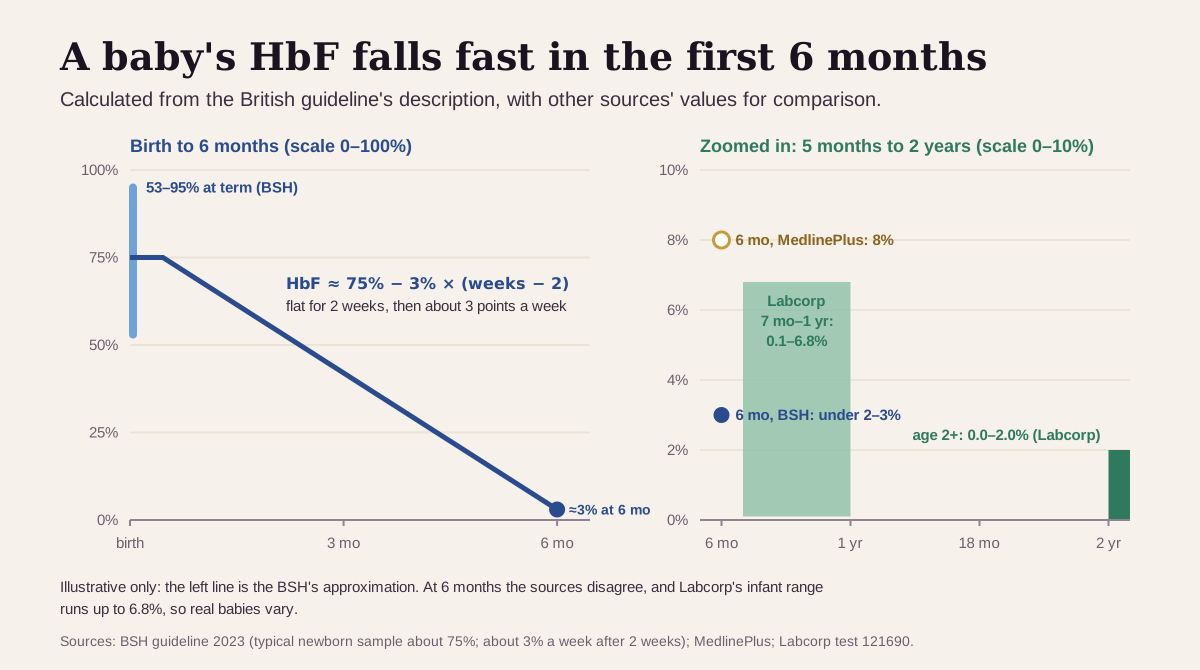
- The first 6 months. HbF holds steady for about 2 weeks, then falls roughly 3 percentage points a week, reaching under 2–3% by 6 months in the BSH's account. Sick preterm babies can switch later.3 Muncie and Campbell describe HbF as "negligible" by about 6 months,9 while MedlinePlus lists 8% at that age.5 Because every newborn has so much HbF, newborn screening can't pick up HPFH or beta-thalassemia trait. Those show up on later testing.3
- Adults. HbF level is largely inherited: its heritability is estimated at about 80%.1 It moves when something changes red-cell production, such as pregnancy (F cells peak at 18–22 weeks)16 or hydroxyurea. In the hydroxyurea trial, the benefits took several months to show,15 and F cells rose over the first year.7
- Transfusions blur the result. The BSH warns that a blood transfusion in the past 4 months can give misleading hemoglobin results,3 and Labcorp asks for transfusion history on the order.2
- Compare like with like. Methods differ most at high levels: HPLC can underestimate a high HbF, while capillary electrophoresis counts modified forms of HbF that HPLC leaves out.3 A trend is easiest to read from the same lab.
Going deeper: the gamma-globin switch
The beta-like globin genes sit together on chromosome 11, and they turn on in a set order through development: an embryonic gene first, then the gamma genes that make HbF, then beta for adult HbA.1 A protein called BCL11A helps keep the gamma genes quiet in adult red-cell precursors. When researchers knocked BCL11A down in the lab, gamma-globin came back on strongly.1
Why HbF differs between healthy adults comes down mostly to genes. Three regions matter most: BCL11A on chromosome 2, the stretch between HBS1L and MYB on chromosome 6, and the beta-globin cluster itself. A 2009 review put their share of the variation at 20–50%, and later, finer mapping at more than 50%.181 High-HbF levels go with less illness and longer survival in both sickle cell disease and beta thalassemia.18
Deletions inside the cluster explain the two high-HbF traits. In delta-beta thalassemia, HbF rises modestly and the globin chains stay somewhat out of balance. In HPFH, HbF rises strongly and chain production stays balanced,1 which fits with HPFH carriers' usually normal red-cell indices.6
The switch is also the target of newer treatments. Exagamglogene autotemcel (exa-cel), approved by the FDA in December 2023, edits a patient's own blood stem cells at the red-cell enhancer of BCL11A to turn HbF production back on.1920 In its main trial, 29 of 30 people with severe sickle cell disease who were followed long enough had no pain crises for at least 12 months.20 Hydroxyurea, an oral medicine approved in 1998, is the older way of raising HbF.1915
Questions worth asking
- Is my HbF normal for my age, and what do my HbA2, HbA and MCV show next to it?
- Does my pattern look more like HPFH, delta-beta thalassemia trait or beta-thalassemia trait, and would DNA testing settle it?
- Have I had a blood transfusion in the past 4 months that could affect this result?
- If I'm pregnant or planning a pregnancy, should my partner be tested too?
- If I have sickle cell disease, what's my HbF now, and how has it changed with my treatment?
- Was this measured by HPLC or capillary electrophoresis, and will my next test use the same lab?
FAQ
What is a normal fetal hemoglobin level? From age 2, Labcorp's range is 0.0–2.0%, and MedlinePlus lists 0.8–2% for adults.25 Newborns are very different: 53–95% at birth, falling over the first year.3
Is a high fetal hemoglobin bad? Usually it's a sign of an inherited trait. HPFH is harmless, delta-beta and beta-thalassemia traits are carrier states, and in sickle cell disease a higher HbF is protective.634
Why does fetal hemoglobin help in sickle cell disease? HbF gets in the way of sickling.15 In one study, levels above about 10% went with fewer strokes and organ complications, and above 20% with fewer pain crises.4 That's why hydroxyurea and gene therapy aim to raise it.1920
Is the fetal hemoglobin test the same as the Kleihauer-Betke test? No. Your HbF result measures your own hemoglobin. The Kleihauer-Betke test looks for a baby's red cells in a mother's blood.8
This guide is for education. It can't diagnose you or replace care from a clinician who knows your history.
Footnotes
-
Sankaran VG, Orkin SH. The switch from fetal to adult hemoglobin. Cold Spring Harb Perspect Med. 2013;3(1):a011643. A review of how the gamma-to-beta switch works: BCL11A, the heritability of HbF (about 80%), and deletional HPFH versus delta-beta thalassemia. PMC ↩ ↩2 ↩3 ↩4 ↩5 ↩6 ↩7 ↩8 ↩9 ↩10 ↩11
-
Labcorp, a commercial lab. Hemoglobinopathy Fractionation Cascade, test 121690. Methods (capillary electrophoresis first, then HPLC) and age-specific reference ranges: Hb F 0.1–6.8% at 7 months to 1 year and 0.0–2.0% from age 2, none established under 7 months. labcorp.com ↩ ↩2 ↩3 ↩4 ↩5 ↩6 ↩7 ↩8 ↩9 ↩10 ↩11 ↩12 ↩13 ↩14 ↩15 ↩16
-
Bain BJ, Daniel Y, Henthorn J, et al. Significant haemoglobinopathies: A guideline for screening and diagnosis: A British Society for Haematology Guideline. Br J Haematol. 2023;201(6):1047–1065. The UK hematology society's guideline. The source for HbF before and after birth (90–95% at 34–36 weeks, 53–95% at term, about 3 points a week down, under 2–3% by 6 months), the 5% and 10% points to look closer, the conditions that call for HbF measurement, method pitfalls and the 4-month transfusion window. doi.org ↩ ↩2 ↩3 ↩4 ↩5 ↩6 ↩7 ↩8 ↩9 ↩10 ↩11 ↩12 ↩13 ↩14 ↩15 ↩16 ↩17 ↩18 ↩19 ↩20 ↩21 ↩22 ↩23 ↩24 ↩25 ↩26 ↩27 ↩28 ↩29 ↩30 ↩31
-
Powars DR, Weiss JN, Chan LS, Schroeder WA. Is there a threshold level of fetal hemoglobin that ameliorates morbidity in sickle cell anemia? Blood. 1984;63(4):921–926. 272 patients over 3,011 patient-years; HbF 2–32% (mean 10%); thresholds of about 10% and 20%. PubMed ↩ ↩2 ↩3 ↩4 ↩5 ↩6 ↩7 ↩8 ↩9 ↩10 ↩11
-
National Library of Medicine. Hemoglobin electrophoresis. MedlinePlus Medical Encyclopedia. Reviewed February 27, 2026. Adult HbF 0.8–2%; newborn 50–80%, 8% at 6 months, 1–2% after 6 months. MedlinePlus ↩ ↩2 ↩3 ↩4 ↩5 ↩6 ↩7 ↩8 ↩9
-
Abdul Hamid FS, Md Noor S, Lai MI, et al. Molecular spectrum and carrier frequency of deletional hereditary persistence of fetal hemoglobin and delta-beta thalassemia in Malaysia. Blood Res. 2025;60(1):51. Gives the typical ranges (HbF 5–15% in delta-beta thalassemia trait, 15–30% in HPFH, citing a standard hemoglobin-disorders text). In its own 534 carriers, delta-beta groups averaged about 14–21% HbF by HPLC, HbA2 was normal or slightly low in every group, and some HPFH groups had mildly small red cells. PMC ↩ ↩2 ↩3 ↩4 ↩5 ↩6 ↩7 ↩8 ↩9 ↩10
-
Steinberg MH, Lu ZH, Barton FB, et al. Fetal hemoglobin in sickle cell anemia: determinants of response to hydroxyurea. Blood. 1997;89(3):1078–1088. HbF after 2 years of hydroxyurea in the same trial, by response quartile. PubMed ↩ ↩2 ↩3 ↩4 ↩5 ↩6
-
Kim YA, Makar RS. Detection of fetomaternal hemorrhage. Am J Hematol. 2012;87(4):417–423. How the Kleihauer-Betke test and anti-HbF flow cytometry work, and their limits. PubMed ↩ ↩2 ↩3 ↩4
-
Muncie HL Jr, Campbell J. Alpha and beta thalassemia. Am Fam Physician. 2009;80(4):339–344. The chain makeup of HbF, HbA and HbA2, HbF about 80% at birth, and the beta-thalassemia trait pattern (MCV usually under 75 fL; raised HbA2, reduced HbA, "probably increased" HbF). PubMed ↩ ↩2 ↩3 ↩4 ↩5 ↩6 ↩7
-
Wood WG, Stamatoyannopoulos G, Lim G, Nute PE. F-cells in the adult: normal values and levels in individuals with hereditary and acquired elevations of Hb F. Blood. 1975;46(5):671–682. F cells average 2.7% (range 0.5–7.0%) of red cells in healthy adults. PubMed ↩ ↩2 ↩3
-
Rodriguez-Capote K, Higgins TN. Incidence of hemoglobinopathies and thalassemias in Northern Alberta. Establishment of reference intervals for HbF and HbA2. Clin Biochem. 2015;48(10–11):698–702. An HbF reference interval of 0.0–1.8% in 3,306 people aged 3–92, plus a year of trait counts. PubMed ↩ ↩2 ↩3 ↩4 ↩5
-
National Heart, Lung, and Blood Institute. Sickle Cell Disease: Symptoms. Updated August 20, 2024. When to seek emergency care. nhlbi.nih.gov ↩ ↩2
-
Platt OS, Brambilla DJ, Rosse WF, et al. Mortality in sickle cell disease. Life expectancy and risk factors for early death. N Engl J Med. 1994;330(23):1639–1644. 3,764 patients; a low HbF was a risk factor for early death in adults with sickle cell anemia. PubMed ↩
-
Platt OS, Thorington BD, Brambilla DJ, et al. Pain in sickle cell disease. Rates and risk factors. N Engl J Med. 1991;325(1):11–16. 18,356 patient-years; high pain rates went with low HbF. PubMed ↩
-
Charache S, Terrin ML, Moore RD, et al. Effect of hydroxyurea on the frequency of painful crises in sickle cell anemia. N Engl J Med. 1995;332(20):1317–1322. The randomized Multicenter Study of Hydroxyurea in 299 adults. PubMed ↩ ↩2 ↩3 ↩4
-
Popat N, Wood WG, Weatherall DJ, Turnbull AC. Pattern of maternal F-cell production during pregnancy. Lancet. 1977;2(8034):377–379. F cells rose in all 11 pregnancies studied, peaking at 18–22 weeks. PubMed ↩ ↩2 ↩3 ↩4
-
Wethers DL. Sickle cell disease in childhood: Part I. Laboratory diagnosis, pathophysiology and health maintenance. Am Fam Physician. 2000;62(5):1013–1020. A table of typical lab patterns in sickle cell disease, and the note that people with sickle cell trait have more than 50% normal hemoglobin. PubMed ↩ ↩2
-
Thein SL, Menzel S. Discovering the genetics underlying foetal haemoglobin production in adults. Br J Haematol. 2009;145(4):455–467. Three gene regions explain 20–50% of HbF variation; high HbF goes with less illness and death in sickle cell disease and beta thalassemia. PubMed ↩ ↩2 ↩3 ↩4
-
National Heart, Lung, and Blood Institute. Sickle Cell Disease: Treatment. Updated September 30, 2024. Hydroxyurea (approved in 1998) and the December 2023 gene-therapy approvals. nhlbi.nih.gov ↩ ↩2 ↩3
-
Frangoul H, Locatelli F, Sharma A, et al. Exagamglogene autotemcel for severe sickle cell disease. N Engl J Med. 2024;390(18):1649–1662. The phase 3 trial of the BCL11A-editing gene therapy. PubMed ↩ ↩2 ↩3
Educational content, not medical advice. Libby is a personal record tool, not a medical service — it doesn't diagnose, treat, or prescribe. Reference ranges vary by lab and by person. Talk to a qualified healthcare professional about your results.
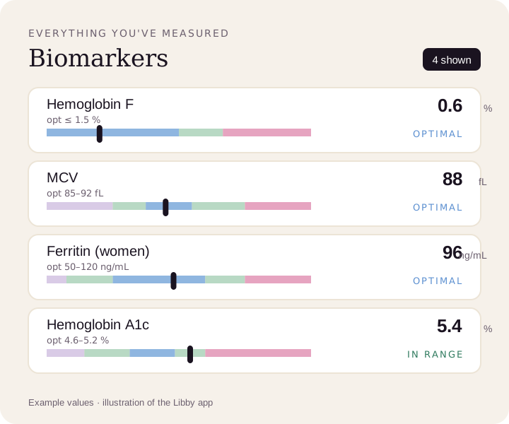
Compare your results to optimal ranges.
Upload a lab PDF or a photo of your report. Libby puts every marker on a reference range and, where the app has one, an optimal range, and tracks your trends over time.
View your hemoglobin F ›