Epithelial cells in urine: what squamous and renal mean
Squamous cells in urine usually mean skin cells got into the sample. Here's what's normal, when renal tubular cells matter, and when to get care.
Epithelial cells are the cells that line your urinary tract and cover your skin, and a few of them in a urine sample are normal. Squamous cells are flat skin-type cells that wash in from the genital skin and, in women, the vagina. Many of them usually means the sample picked up cells from outside the urinary tract. The type that matters most is the renal tubular cell, which comes from inside the kidney and points to kidney injury.123
The short version
- Normal: "few" cells. Labcorp's reference interval for non-renal epithelial cells is 0–10 per high-power field (hpf). Renal tubular cells should be absent.142
- Many squamous cells usually means a contaminated sample: skin or vaginal cells, and their bacteria, got into the cup. In one emergency department, 52% of urine samples had more than 5 per hpf. The usual fix is a new midstream clean-catch sample.526
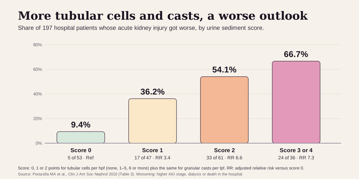
- Renal tubular cells point to injury of the kidney's tiny tubes, most often acute tubular necrosis. In hospital patients with kidney injury, 9% with no tubular cells or casts got worse, versus 67% with the most.27
- Get care now if you make little or no urine, or have new swelling in your legs or feet, confusion, or nausea and vomiting. Those are signs of acute kidney failure.89
What epithelial cells are
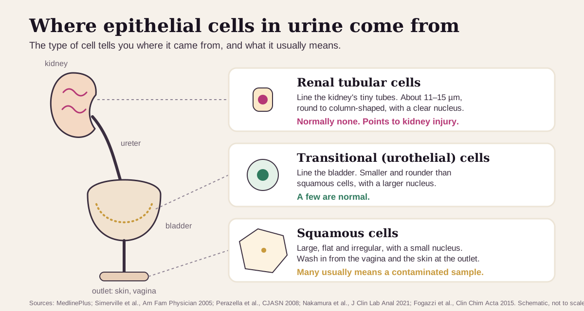
Epithelial cells cover the inside and outside surfaces of your body, including your kidneys, ureters, bladder and urethra. Three types can show up in urine:1
- Squamous cells are large, flat and irregular, with a small nucleus. In a urine sample, their presence suggests contamination, most often from the vagina or the skin around the urethra.23
- Transitional cells, also called urothelial cells, line the bladder. They're smaller and rounder than squamous cells, with larger nuclei, and a few of them are normal.211
- Renal tubular cells line the tiny tubes inside the kidney. They're about 11–15 µm across, round to column-shaped, with a clear nucleus. Their presence indicates real kidney disease.102
The count comes from the microscopic part of a urinalysis. A lab spins down the urine and looks at the sediment under the microscope.21 Labcorp's report prints two lines: Epithelial Cells (non renal) and Epithelial Cells (renal).4
What's a normal epithelial cell count?
"Few" is normal, according to MedlinePlus.1 In numbers:
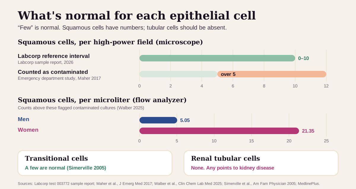
- Squamous (non-renal) cells: Labcorp's reference interval is 0–10 per hpf.4 Researchers studying contamination draw the line lower. One emergency department study counted more than 5 per hpf as a contaminated sample.5
- Transitional cells: a few are normal.2
- Renal tubular cells: none. Their presence indicates significant kidney disease.2
Women's limit is higher than men's. In 2,527 samples run on a flow analyzer, the counts that flagged a contaminated culture were 21.35 squamous cells per µL for women and 5.05 per µL for men, about four times higher.12
"Optimal" squamous cells: fewer means a cleaner sample
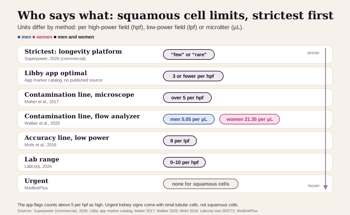
For squamous cells, "optimal" is about the sample more than about you: the fewer skin cells in the cup, the more you can trust the rest of the urinalysis.13 The Libby app shows 3 or fewer per hpf as optimal. Here's the full ladder, from strictest to loosest:
| Line | Men | Women | Who uses it |
|---|---|---|---|
| Strictest (commercial) | "few" or "rare" | "few" or "rare" | Superpower's published optimal14 |
| Libby app optimal | 3 or fewer per hpf | 3 or fewer per hpf | The optimal range shown in the Libby app. The app's catalog records no published source for this band, and the app flags counts above 5 as high |
| Contamination line (microscope) | over 5 per hpf | over 5 per hpf | An emergency department study's definition of a contaminated sample5 |
| Contamination line (flow analyzer) | 5.05 per µL | 21.35 per µL | Walber et al., the counts that flagged mixed-growth cultures12 |
| Accuracy line (low power) | 8 per lpf | 8 per lpf | Mohr et al.: above this, the urinalysis predicted bacteria in urine less well13 |
| Lab range | 0–10 per hpf | 0–10 per hpf | Labcorp's reference interval4 |
| Urgent | none for squamous cells | none for squamous cells | Urgent kidney signs come with renal tubular cells (below)8 |
Why fewer squamous cells matters:
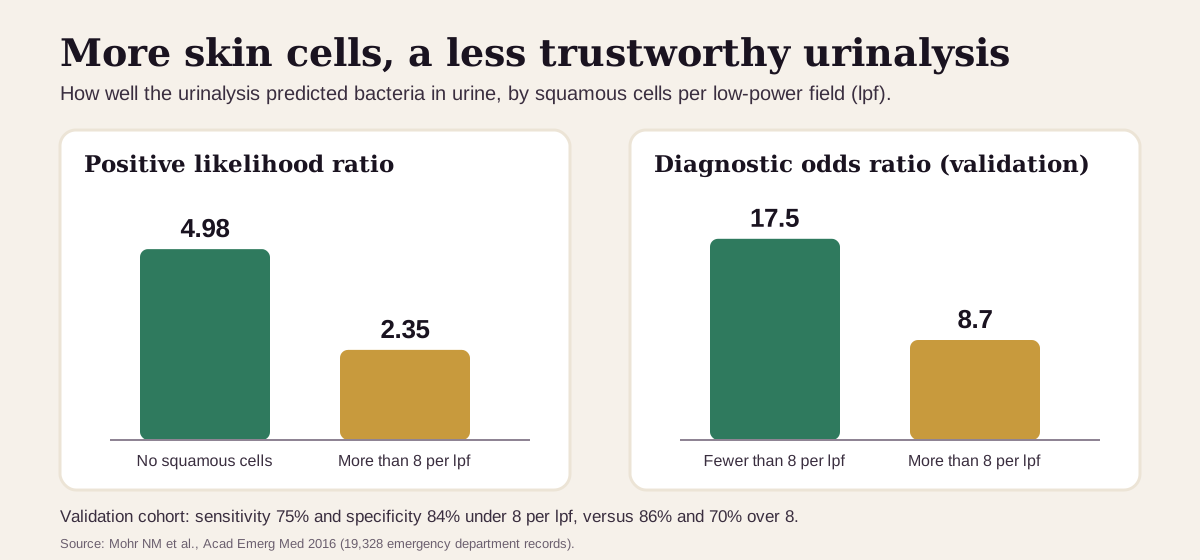
- In 19,328 emergency department urinalyses, the standard urine signs of bacteria were much more telling with no squamous cells than with more than 8 per lpf. The positive likelihood ratio fell from 4.98 to 2.35. In a separate check, samples under 8 per lpf gave a diagnostic odds ratio of 17.5, versus 8.7 above it.13
- In 2,527 hospital samples, a squamous count above the sex-specific cutoff made a mixed-growth (contaminated) culture 2.78 times as likely.12
Keep "optimal" in perspective: a low squamous count is a quality mark, not a health score. The same study found squamous cells were a poor predictor of a contaminated culture (area under the curve 0.68), so a clean-looking sample can still be contaminated.13 Technique matters, but less than you'd think. A review of the evidence for the CDC found that wiping first made no difference to contamination in women, while a midstream sample cut contamination in men compared with the first stream.6
What a high count usually means
Start with the cell type. A high squamous count is usually about how the sample was collected. Transitional cells are usually harmless in small numbers. Renal tubular cells are the ones that point inside the kidney.21
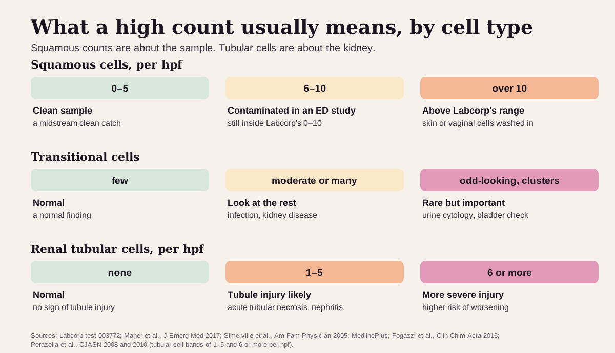
Many squamous cells: usually the sample
This is by far the most common reason. Urine can easily pick up bacteria from the skin around the urethra, the vagina and the anus on its way into the cup.6 Squamous cells are the visible sign of that, so a sample with many of them can also show bacteria that came from outside the urinary tract, and the rest of the urinalysis gets less reliable.133 Contamination is also a known cause of a false-positive white-cell (leukocyte esterase) dipstick.2
What usually happens next is a repeat clean-catch sample. MedlinePlus describes the steps:151
- Wash your hands. With a vagina, separate the labia and wipe front to back. With a penis, wipe the head, pulling back the foreskin first.
- Start peeing into the toilet, stop, then catch the middle of the stream in the cup without letting it touch your body.
- Tell the clinic if you're on your period.
Even so, contamination is common. In one emergency department, posting written instructions left the contaminated share unchanged at 52%.5
Transitional cells: a few are normal
A few transitional cells are a normal finding.2 MedlinePlus lists urinary tract infection, kidney disease and certain cancers among the causes of "moderate" or "many" epithelial cells.1 Rarely, the cells look abnormal: unusual sizes and shapes, large nuclei, several nuclei, often in clusters. One report found bladder cancer by cystoscopy in all three patients whose routine urine sediment showed cells like these. Urine cytology, a stained look at the cells in a pathology lab, is the standard test for them.11
Renal tubular cells: a kidney signal
Their presence indicates significant kidney disease.2 The usual cause comes first, then the less common ones:
- Acute tubular necrosis (ATN), injury to the kidney's tubule cells, is a common cause of kidney failure in people in the hospital. It usually follows a drop in blood flow: low blood pressure for more than 30 minutes, septic shock, major surgery, or muscle injury. Some medicines damage the tubules directly, such as aminoglycoside antibiotics and the antifungal amphotericin, and so can contrast dye.9
- Other kidney conditions that shed tubular cells into casts include interstitial nephritis, eclampsia, transplant rejection and heavy-metal poisoning.2
- Heavy protein leak (nephrotic syndrome) fills tubular cells with fat. The lab calls them oval fat bodies, or fatty casts when they're packed into a cast.24
Tubular cells help tell ATN from a milder kidney dip. In 267 hospital patients with kidney injury, when clinicians already suspected ATN, finding any tubular cells or granular casts confirmed it every time (positive predictive value 100%). When they suspected a milder, fluid-related cause, finding none made ATN unlikely (negative predictive value 91%).10
The more tubular cells, the worse the outlook. In a second study of 197 patients, the chance that kidney injury got worse in the hospital rose with a score built from tubular cells and granular casts:7
Get care now if you're making little or no urine, or if a kidney finding comes with new swelling in your legs, ankles or feet, confusion or drowsiness, nausea and vomiting, or shortness of breath. Those can be signs of acute kidney failure.89
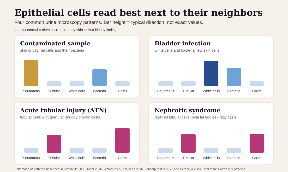
Epithelial cells read best next to their neighbors
The cell type tells you where to look. The markers around it tell you what's going on:
| If you see… | It often points to… |
|---|---|
| Many squamous cells, often with mixed bacteria | a contaminated sample: skin or vaginal cells and their bacteria got into the cup. Repeat it as a clean catch |
| White cells and bacteria, few squamous cells, no casts | a bladder infection, especially with burning or frequent urination1 |
| Renal tubular cells with granular ("muddy brown") casts | acute tubular necrosis, especially in someone who's been in the hospital |
| Oval fat bodies or fatty casts | nephrotic syndrome, a heavy protein leak from the kidney's filters |
| Odd-looking transitional cells, sometimes in clusters | a reason for urine cytology and a bladder check11 |
Casts form inside the kidney. Apart from a few clear (hyaline) casts, which can be normal after exercise, finding them moves the question from the bladder to the kidney itself.24
How fast it changes
A squamous count belongs to one sample. It reflects how that sample was collected, so a new clean-catch sample gives a fresh reading.115
Timing changes the result. Urine should be looked at within 2 hours of collection; longer delays often give unreliable results.2 Left at room temperature for more than 4 hours, both real and contaminating bacteria overgrow. Refrigeration or a boric acid preservative holds a sample for up to 24 hours.6
Renal tubular cells track the kidney injury. ATN can last from a few days to 6 weeks or more, and kidney function often returns to normal.9 At the time of a kidney consult, the sediment score predicted worsening better than the kidney-injury stage did.7
Going deeper: telling the three cells apart
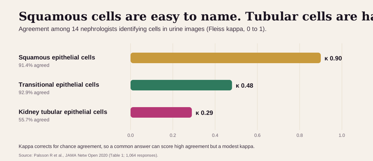
Squamous cells are easy to spot; tubular cells are hard. When 14 nephrologists identified cells in urine images, they agreed on squamous cells almost every time (kappa 0.90). Agreement fell to 0.48 for transitional cells and 0.29 for kidney tubular cells, with only 55.7% agreeing on tubular cells.16
Machines count them differently. Automated urine analyzers count red and white cells accurately, but they're less reliable for kidney tubular cells and casts, which are best judged by a person at the microscope.17 One analyzer sorts epithelial cells into "squamous" and "nonsquamous".3 Flow analyzers report cells per µL rather than per hpf, which is why the research cutoffs above use different units.12
The sediment score is simple arithmetic. Perazella's group gave 0, 1 or 2 points for tubular cells (none, 1–5, or 6 or more per hpf) and the same for granular casts (per lpf), for a total of 0 to 4.7 In their earlier study, finding any tubular cells or granular casts was the strong ATN signal.10
Questions worth asking
- Were these squamous cells, transitional cells or renal tubular cells?
- If squamous cells were high, should I repeat the test as a clean catch?
- Do the white cells, bacteria or culture still count if the sample was contaminated?
- If renal tubular cells were seen, what do my creatinine and urine protein show?
- Were there casts, and what kind?
- If the transitional cells looked unusual, do I need urine cytology or a bladder check?
FAQ
What does it mean to have squamous epithelial cells in urine? Usually that skin or vaginal cells got into the sample. Labcorp's range for non-renal epithelial cells is 0–10 per hpf, and the midstream clean-catch method is how you keep them out.421
How many epithelial cells in urine is normal? "Few" is normal. Labcorp's reference interval is 0–10 per hpf for non-renal cells, and renal tubular cells should be absent.142
Are renal tubular epithelial cells serious? They indicate kidney disease, most often injury to the kidney's tubules (acute tubular necrosis) in people who are in the hospital. The more there are, the more likely the injury is to worsen.27
Can a contaminated urine sample look like a UTI? Yes. Urine can pick up bacteria from the skin and vagina, and with more than 8 squamous cells per lpf the usual urine signs of bacteria were far less accurate.613
This guide is for education. It can't diagnose you or replace care from a clinician who knows your history.
Footnotes
-
National Library of Medicine. Epithelial Cells in Urine. MedlinePlus. Last updated November 12, 2024. The three cell types, "few" as a normal result, the causes of "moderate" or "many", and squamous cells as a sign of a contaminated sample. MedlinePlus ↩ ↩2 ↩3 ↩4 ↩5 ↩6 ↩7 ↩8 ↩9 ↩10 ↩11 ↩12 ↩13 ↩14 ↩15
-
Simerville JA, Maxted WC, Pahira JJ. Urinalysis: a comprehensive review. Am Fam Physician. 2005;71(6):1153–1162. What each cell looks like and means, the cast table, and the two-hour rule. AAFP ↩ ↩2 ↩3 ↩4 ↩5 ↩6 ↩7 ↩8 ↩9 ↩10 ↩11 ↩12 ↩13 ↩14 ↩15 ↩16 ↩17 ↩18 ↩19 ↩20 ↩21 ↩22 ↩23 ↩24 ↩25
-
Nakamura A, Shinke T, Noguchi N, et al. Evaluation of the detection ability of uropathogen morphology and vaginal contamination by the Atellica UAS800 automated urine microscopy analyzer and its effectiveness. J Clin Lab Anal. 2021;35(3):e23698. Squamous epithelial cells as an indicator of vaginal contamination. PMC ↩ ↩2 ↩3 ↩4 ↩5
-
Labcorp. Urinalysis, Complete With Microscopic Examination (test 003772), test page and sample report (date reported May 18, 2026). Reference interval for Epithelial Cells (non renal): 0–10 /hpf; notes on renal tubular and fatty casts. A national commercial lab's published range. Labcorp ↩ ↩2 ↩3 ↩4 ↩5 ↩6 ↩7 ↩8 ↩9 ↩10 ↩11 ↩12
-
Maher PJ, Brown AEC, Gatewood MO. The Effect of Written Posted Instructions on Collection of Clean-Catch Urine Specimens in the Emergency Department. J Emerg Med. 2017;52(5):639–644. Contamination defined as more than 5 squamous cells per hpf; 392 of 754 urinalyses (52%) contaminated. DOI ↩ ↩2 ↩3 ↩4 ↩5 ↩6 ↩7
-
LaRocco MT, Franek J, Leibach EK, et al. Effectiveness of Preanalytic Practices on Contamination and Diagnostic Accuracy of Urine Cultures: a Laboratory Medicine Best Practices Systematic Review and Meta-analysis. Clin Microbiol Rev. 2016;29(1):105–147. The CDC Laboratory Medicine Best Practices review of urine collection and storage. PMC ↩ ↩2 ↩3 ↩4 ↩5 ↩6
-
Perazella MA, Coca SG, Hall IE, et al. Urine microscopy is associated with severity and worsening of acute kidney injury in hospitalized patients. Clin J Am Soc Nephrol. 2010;5(3):402–408. 197 patients with ATN or prerenal injury; worsening in 9.4%, 36.2%, 54.1% and 66.7% by score. PMC ↩ ↩2 ↩3 ↩4 ↩5 ↩6
-
National Library of Medicine. Acute kidney failure. MedlinePlus Medical Encyclopedia. Review date May 6, 2024. Symptoms, and when to contact a provider. MedlinePlus ↩ ↩2 ↩3
-
National Library of Medicine. Acute tubular necrosis. MedlinePlus Medical Encyclopedia. Review date September 2, 2025. Causes, symptoms and how long ATN lasts. MedlinePlus ↩ ↩2 ↩3 ↩4
-
Perazella MA, Coca SG, Kanbay M, et al. Diagnostic value of urine microscopy for differential diagnosis of acute kidney injury in hospitalized patients. Clin J Am Soc Nephrol. 2008;3(6):1615–1619. 267 patients; tubular cells 11–15 µm; odds ratio 74 and negative predictive value 91%. PMC ↩ ↩2 ↩3 ↩4 ↩5 ↩6
-
Fogazzi GB, Pallotti F, Garigali G. Atypical/malignant urothelial cells in routine urinary sediment: worth knowing and reporting. Clin Chim Acta. 2015;439:107–111. Three patients with bladder carcinoma found after abnormal urothelial cells in routine sediment; cytology as the gold standard. PubMed ↩ ↩2 ↩3 ↩4 ↩5
-
Walber GB, Poloni JAT, Rotta LN. Quality indicators for urine sample contamination: can squamous epithelial cells and bacteria count be used to identify properly collected samples? Clin Chem Lab Med. 2025;63(5):979–986. 2,527 samples on a Sysmex UF-5000 flow analyzer. DOI ↩ ↩2 ↩3 ↩4 ↩5 ↩6 ↩7
-
Mohr NM, Harland KK, Crabb V, et al. Urinary Squamous Epithelial Cells Do Not Accurately Predict Urine Culture Contamination, but May Predict Urinalysis Performance in Predicting Bacteriuria. Acad Emerg Med. 2016;23(3):323–330. 19,328 emergency department records. DOI ↩ ↩2 ↩3 ↩4 ↩5 ↩6 ↩7 ↩8 ↩9
-
Superpower. Urine Epithelial Cells: Best Biomarkers. Last updated September 1, 2026. States that "optimal results typically fall in the 'few' or 'rare' category." A commercial source, listed as what an optimizer platform says. superpower.com ↩ ↩2
-
National Library of Medicine. Clean catch urine sample. MedlinePlus Medical Encyclopedia. Review date July 23, 2024. MedlinePlus ↩ ↩2
-
Palsson R, Colona MR, Hoenig MP, et al. Assessment of Interobserver Reliability of Nephrologist Examination of Urine Sediment. JAMA Netw Open. 2020;3(8):e2013959. 14 nephrologists, 1,064 interpretations. PMC ↩ ↩2
-
Hoenig MP, Mena JD, Lecker SH. More than meets the I(ris): Use of manual urine microscopy to complement automated findings in acute kidney injury. Pract Lab Med. 2022;29:e00267. What automated analyzers count well and what needs a microscope. PMC ↩
Educational content, not medical advice. Libby is a personal record tool, not a medical service — it doesn't diagnose, treat, or prescribe. Reference ranges vary by lab and by person. Talk to a qualified healthcare professional about your results.
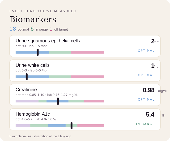
See your urinalysis on your lab's range and the optimal range.
Upload a lab PDF or a photo of your report. Libby puts every marker on your lab's range, adds the optimal range where one exists, and tracks each urinalysis over time.
View your epithelial cells ›