Red blood cells in urine: what your RBC/hpf count means
Urine red cells are counted per high-power field under a microscope. What's normal, what misshapen cells and casts mean, and the AUA/SUFU workup.
The urine red-cell count is the number of red blood cells a lab sees in one microscope field of your spun-down urine. Healthy urine has almost none: 0 to 2 per high-power field (hpf). A count of 3 or more is called microscopic hematuria, and the shape of the cells helps show where they came from: misshapen cells and red-cell "casts" point to the kidney's filters, while round, normal-looking cells point lower down, to the bladder, prostate or a stone.1234
The short version
- Normal: 0–2 red cells per high-power field (RBC/hpf) on a Labcorp report.1 The U.S. urology guideline (AUA/SUFU) calls 3 or more on one properly collected sample microhematuria.2
- The usual reasons for a confirmed high count are a urinary tract infection, an enlarged prostate and kidney stones. In 43–68% of people, no cause turns up.5 A hard workout, a period, sex or a recent catheter can raise it for a short time.54
- The shape is the key split. Misshapen (dysmorphic) red cells, red-cell casts and protein point to the kidney's filters (glomerular disease). Round red cells with no casts point to bleeding lower in the urinary tract.34
- Get care right away for visible blood with clots, trouble passing urine, or fever with pain in your side or back.63
What the urine red-cell count is
The count comes from the microscope part of a urinalysis. A lab spins about 10 mL of fresh urine, pours off the top, and puts a drop of the sediment under a microscope. A technologist reads at least 10–20 fields at 400×, and the result is given as red cells per field.5 Some labs report a range instead of a single number: a Labcorp sample report shows a result of "11-30" per hpf.1
Some labs use automated analyzers that count cells per µL of unspun urine. The American Urological Association (AUA) still treats the microscope count as the reference standard for defining blood in the urine.2
The count usually follows a positive dipstick. The dipstick "blood" pad reacts to heme, and the AUA/SUFU guideline says any positive pad, even a trace, should be checked with a microscope count, which is what defines microhematuria.2
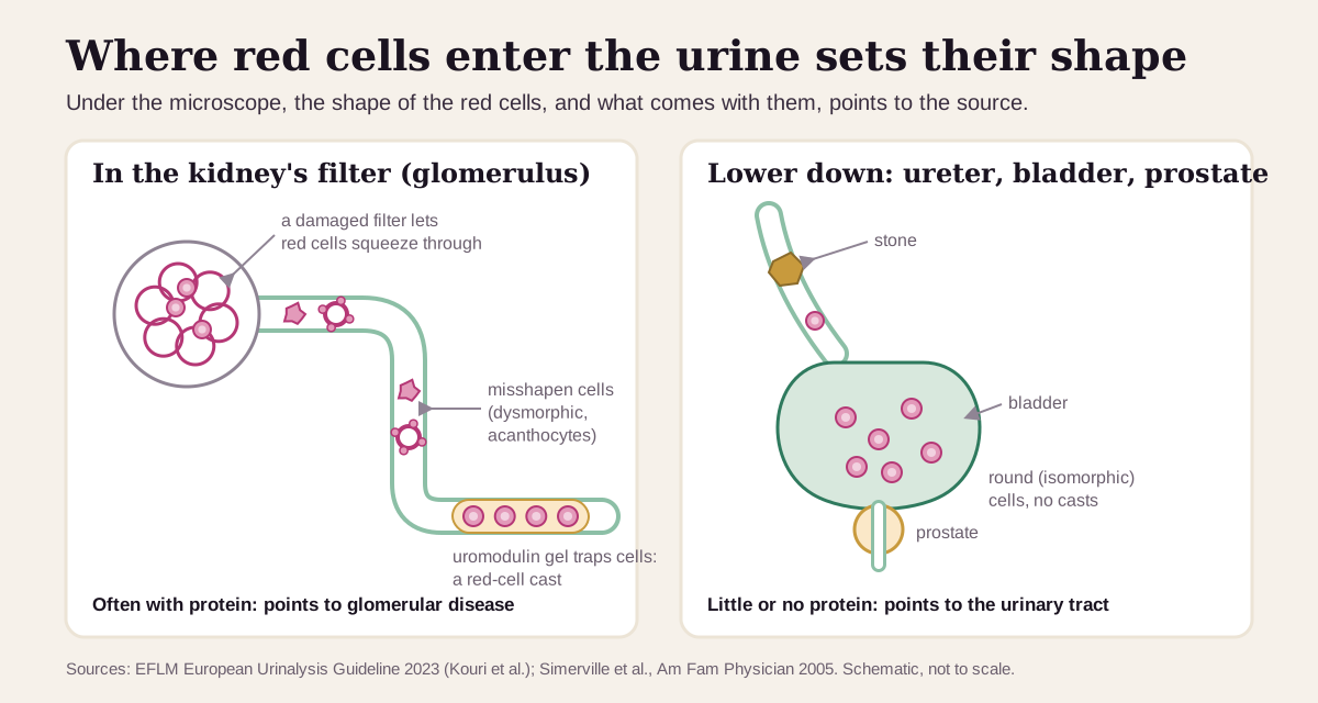
Red cells can enter urine anywhere from the kidney's filters (glomeruli) to the tip of the urethra. Where they enter changes how they look:
- Red cells that squeeze through a damaged kidney filter often come out with odd shapes (dysmorphic).4 The most telling shape is the acanthocyte, a ring-shaped cell with small blebs on its surface.3
- Further down the kidney tubule, a protein called uromodulin (Tamm-Horsfall protein) can set like gel around those cells and form a tube-shaped red-cell cast. Red-cell casts always mean bleeding from the kidney tissue itself.34
- Red cells from the bladder, prostate, a stone or the urethra usually keep their normal round shape (isomorphic).3
(More on the shapes and the counting method at the end.)
What's a normal urine red-cell count?
Normal is 0–2 red cells per high-power field. That's the range printed on a urinalysis report from Labcorp, a commercial lab.1 The AUA/SUFU guideline draws the line just above it: 3 or more RBC/hpf on a single, properly collected sample is microhematuria and starts a workup.2
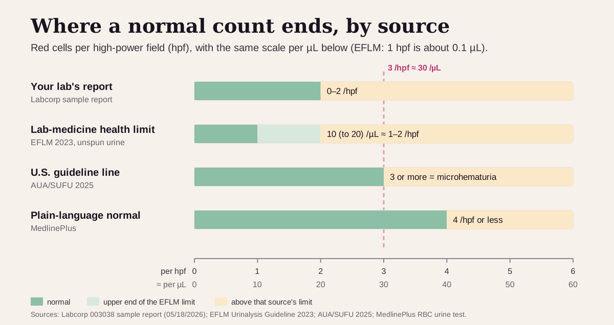
- The European Federation of Clinical Chemistry and Laboratory Medicine (EFLM) puts the healthy upper limit at roughly 10 (to 20) red cells per µL of unspun morning urine, about 1–2 per hpf on its own conversion.3
- MedlinePlus, the U.S. National Library of Medicine's site, gives a looser plain-language line: 4 RBC/hpf or less, adding that ranges vary by lab.7
- Microscopic blood is common. The EFLM puts isolated microscopic blood at 4–13% of the population, mostly from infections or stones, and often below about 30 per µL (about 3 per hpf).3
These cut-offs are the same for men and women. The AUA/SUFU uses sex and age only to sort cancer risk, further down.2
How the sample is collected changes the count. The AUA/SUFU asks for a midstream clean catch: let the first 10 mL go into the toilet, then collect. It advises against testing urine passed right after a night lying down or right after hard exercise or sex, and it asks women to repeat the test after a period ends.2 More than 5 flat squamous (skin) cells per hpf suggests the sample picked up cells on the way out.5 Very dilute urine (below about 308 mOsm) bursts red cells, so the count can read falsely low.2
"Optimal" urine red cells: what health optimizers aim for
Everyone aims for as few red cells as possible. Where sources differ is how many count as "some". Here's the ladder, strictest first:
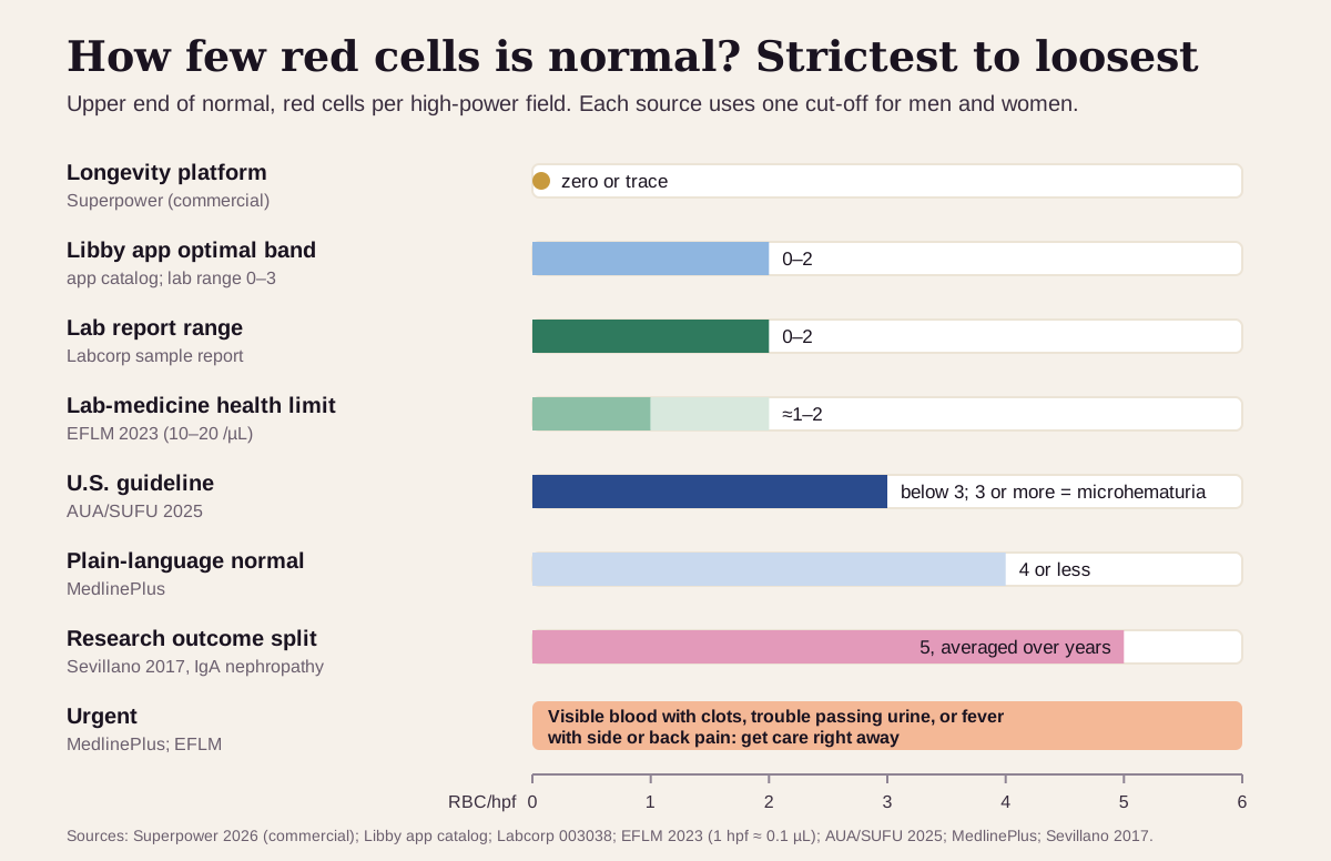
| Target | Red cells per hpf | Who uses it |
|---|---|---|
| Strictest (longevity platform) | zero or trace | Superpower: "Normally, urine contains zero or trace RBCs; any persistent elevation demands investigation"8 |
| Libby app | optimal 0–2; flagged high above 3 | The optimal band shown in the Libby app; its lab range is 0–3 |
| Lab report | 0–2 | Labcorp's printed reference interval1 |
| Lab-medicine health limit | about 1–2 (10–20 per µL, unspun) | The EFLM's European urinalysis guideline3 |
| U.S. guideline | 3 or more = microhematuria | AUA/SUFU 2025, on one properly collected sample2 |
| Plain-language normal | 4 or less | MedlinePlus7 |
| Research outcome split | above 5, averaged over years | In IgA nephropathy, the split between better and worse kidney survival9 |
| Urgent | visible blood with clots, trouble passing urine, or fever with side or back pain | Get care right away63 |
Why the low line matters:
- In a study of more than 46,000 patients cited by the AUA/SUFU, a cut-off of 3–10 RBC/hpf gave the best sensitivity for finding bladder cancer (0.73). The guideline keeps its line low so that fewer cancers slip past.2
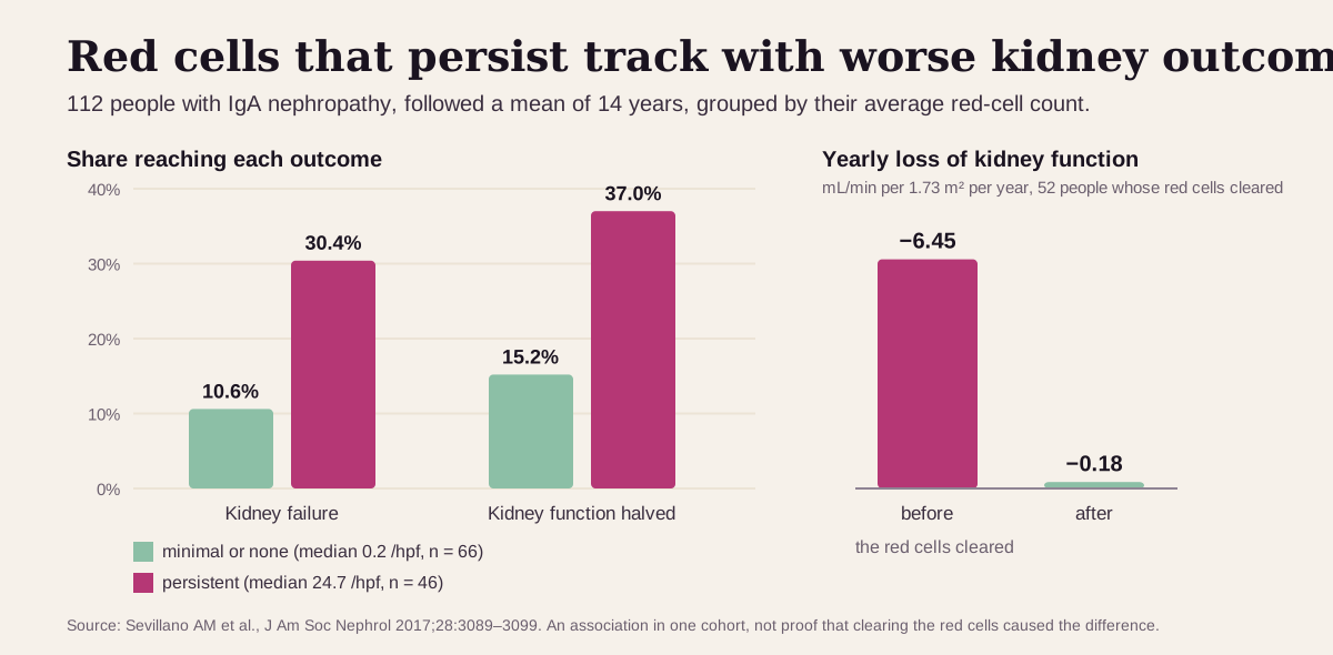
- In 112 people with IgA nephropathy followed for about 14 years, those whose red cells stayed high (averaging about 25 per hpf) reached kidney failure nearly 3 times as often as those whose count stayed near zero: 30.4% vs 10.6%. Losing half their kidney function followed the same pattern: 37.0% vs 15.2%.9
- In the same study, once the red cells disappeared, kidney function decline slowed from 6.45 to 0.18 mL/min per 1.73 m² per year.9
Keep "optimal" in perspective: a count of 3 is a reason to look, not a diagnosis. Most people with microscopic blood have nothing serious, and 43–68% have no cause found at all.45 The EFLM also notes a little "physiological" blood in healthy urine.3 In the lowest-risk group, the AUA/SUFU recommends simply repeating the urinalysis within six months.2 There's no "too low": zero is as good as this count gets.7
What a high urine red-cell count usually means
First, rule out the temporary causes. Clinicians look for these before anything else, then repeat the count:52
- Hard exercise. Long-distance running is the classic example. A repeat urinalysis 48–72 hours later should be clear.4
- A period, sex, a viral illness, a minor injury or a recent catheter. The cause is removed or treated and the urine retested after at least 48 hours.5
- Contamination. Many squamous cells in the sample mean skin or vaginal cells got in. The fix is a new clean-catch sample or, sometimes, a catheter sample.25
If it's confirmed, the usual suspects in people with microscopic blood are, roughly from most to least common:5
- No cause found: 43–68%.
- Urinary tract infection: 4–22%. White cells, burning, and a positive leukocyte or nitrite pad point to it. The AUA/SUFU asks for a repeat microscope count after treatment to confirm the blood has cleared.24
- Enlarged prostate (BPH): 10–13%.
- Kidney stones: 4–5%. Pain in the side (flank) is a clue.2
- Bladder cancer: 2–4%. Risk rises with male sex, age over 35 and smoking.5
- Less common: kidney cysts and kidney disease (about 2–3% each); kidney cancer, prostate cancer and a narrowed urethra (under 1% each).5
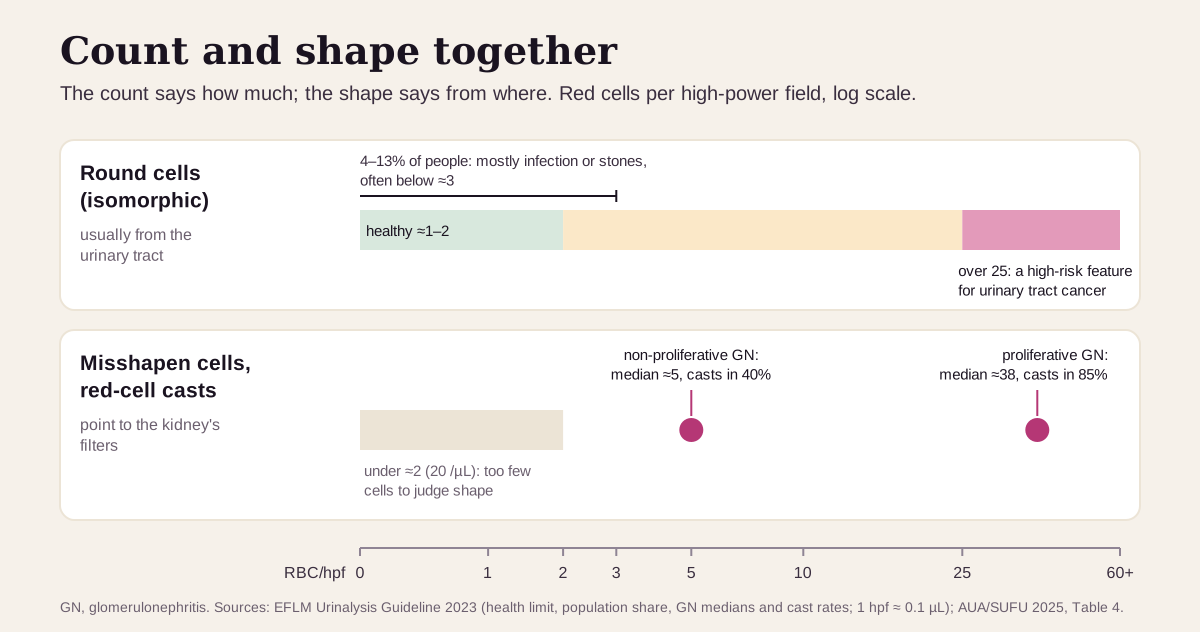
When the cells are misshapen, think kidney. The most common kidney-filter cause is IgA nephropathy, an immune disease of the glomeruli.4 Other kidney causes include Alport syndrome and benign familial hematuria.5 In the EFLM's summary, proliferative (more active) glomerulonephritis ran a median of about 38 RBC/hpf with red-cell casts in 85%, while non-proliferative forms ran about 5 RBC/hpf with casts in 40%.3 About 20% of people with biopsy-proven glomerulonephritis show blood alone, without the other signs.4
Blood thinners such as aspirin or warfarin can cause blood in the urine,6 but the AUA/SUFU asks for the same workup whether or not you take them.2
Get care right away if you pass blood clots, can't urinate, or have fever, shaking chills, nausea or pain in your belly, side or back along with blood in your urine.6 Fever with flank pain can mean a kidney infection, which the EFLM treats as an emergency.3 Dark, rust-colored or brown urine, foamy urine, and swelling of the face, ankles or legs are symptoms of glomerulonephritis; MedlinePlus advises contacting your clinician if they appear.10
What happens after a high count: the AUA/SUFU workup
The AUA/SUFU guideline (2020, amended 2025) sets out the U.S. path once a count of 3 or more is confirmed:2
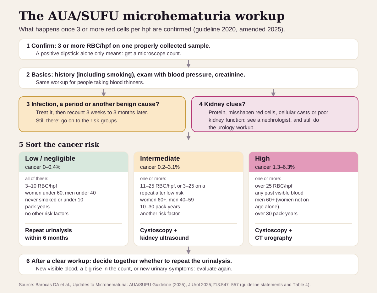
- Confirm it. One properly collected sample with 3 or more RBC/hpf is enough. A positive dipstick alone is only a reason to count.2
- Start with the basics. A history (including smoking), a physical exam with blood pressure, and a creatinine blood test.2
- Treat anything temporary, then recount. After an infection or another benign cause clears, the guideline panel suggests waiting at least 3 weeks and no more than 3 months before repeating the urinalysis. If the blood persists, the risk-based workup starts.2
- Look for kidney clues. Protein, misshapen red cells, cellular casts or poor kidney function point to kidney disease and a referral to a kidney specialist (nephrologist). The urologic workup still goes ahead too.2
- Sort the cancer risk by count, age, sex, smoking and other risk
factors:2
- Low (3–10 RBC/hpf; women under 60, men under 40, fewer than 10 pack-years, and nothing else): cancer in 0–0.4%. Repeat the urinalysis within six months.
- Intermediate (11–25 RBC/hpf, or any one of: women 60 or older, men 40–59, 10–30 pack-years, another risk factor): cancer in 0.2–3.1%. Cystoscopy (a camera look inside the bladder) and a kidney ultrasound.
- High (over 25 RBC/hpf or any past visible blood, or men 60 or older, or more than 30 pack-years): cancer in 1.3–6.3%. Cystoscopy and a CT urogram. Women aren't placed in the high group on age alone.
- After a clear workup, you and your clinician decide together whether to repeat the urinalysis. New visible blood, a big jump in the count, or new urinary symptoms restart the workup.2
Urine red cells read best next to their neighbors
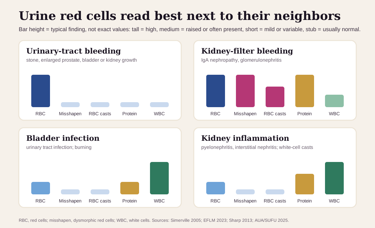
A red-cell count rarely comes alone. The rest of the microscope report and the dipstick usually say more than the count does by itself:34
| If you see… | It often points to… |
|---|---|
| Many round red cells, no protein, no casts, no or only a few white cells | bleeding along the urinary tract: a stone, an enlarged prostate, or a bladder or kidney growth |
| Misshapen red cells (40% or more, or acanthocytes 2% or more), red-cell casts, protein | the kidney's filters (glomerular disease); IgA nephropathy is the most common cause |
| Red cells with many white cells, burning when you pass urine | a bladder infection; the count is repeated after treatment |
| White-cell casts and protein | inflammation of the kidney tissue: a kidney infection (pyelonephritis) or interstitial nephritis. Fever with flank pain points to pyelonephritis and is urgent |
| Red cells with more than 5 squamous cells per hpf, or during a period | contamination; repeat with a clean-catch sample (after the period ends) or a catheter sample |
| A high count within a day or two of a hard workout | exercise; a repeat 48–72 hours later should be clear |
When the kidney-filter pattern shows up, clinicians measure the urine protein (a spot protein-to-creatinine ratio or a 24-hour collection) and kidney function,4 and refer to a nephrologist alongside the urologist.25
How fast the count changes
Red cells in a urine sample fade fast. The sample should be looked at within two hours of collection.4 Red cells break up in dilute urine,2 and spinning the sample can lose 20–80% of them.3 That's one reason two counts from the same day can differ.
In your body, the timing depends on the cause:
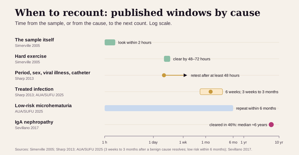
- Exercise: clear on a repeat 48–72 hours later.4
- A period, sex, a viral illness or a catheter: retest after at least 48 hours.5
- An infection: recount about 6 weeks after treatment.5 The AUA/SUFU panel notes that blood can take several weeks to a few months to clear, and suggests recounting between 3 weeks and 3 months.2
- Kidney-filter disease: slow. In the IgA nephropathy study, the red cells disappeared in 46% of people, and that took a median of about 6 years.9
Trends beat snapshots. A single urinalysis catches about 95% of people with microscopic blood, and the guideline treats one positive count as enough to act on.2 After a clear workup, a count that jumps, or new visible blood, restarts the evaluation.2 Comparing results works best from the same lab, since labs use different methods and samples.7
Going deeper: shapes, casts and how the count is made
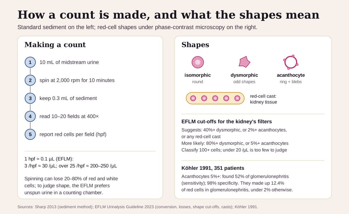
Counting. A standard sediment starts with about 10 mL of midstream urine, spun at 2,000 rpm for 10 minutes. The sediment is resuspended in 0.3 mL and at least 10–20 fields are read at 400×.5 Spinning can lose 20–80% of the cells, so for judging shape the EFLM prefers unspun urine in a counting chamber. It recommends reporting per liter: red cells × 10⁶/L, which equals red cells per µL.3 By its conversion, 3 RBC/hpf is about 30 per µL and 25 RBC/hpf is about 200–250 per µL.3
Shapes. The EFLM recommends phase-contrast microscopy, which shows the outline of each cell, and classifying at least 100 red cells.3 Its cut-offs:3
- Suggests kidney-filter bleeding: 40% or more dysmorphic cells, or 2% or more acanthocytes, or any red-cell cast.
- Makes it more likely: 80% or more dysmorphic cells, or 5% or more acanthocytes.
- Too few cells to judge: under 20 red cells per µL, because a little healthy "physiological" blood can look misshapen too.
Acanthocytes carry the most weight. In a German study of 351 people with blood in the urine, acanthocytes made up 12.4% of red cells in biopsy-proven glomerulonephritis and under 2% in other causes. A cut-off of 5% or more caught 52% of the glomerulonephritis cases (sensitivity) and was wrong in only 4 of 187 people with other causes (specificity 98%).11
Casts. Casts form in the last stretch of the kidney tubule and the collecting ducts, from gelled uromodulin made higher up the tubule.3 Whatever is in the tubule gets trapped: red cells make a red-cell cast, white cells a white-cell cast.4 Red-cell casts mean bleeding from kidney tissue.3 One quirk: they can turn up in people who play contact sports.4
Questions worth asking
- How many red cells per high-power field did the lab count, and was the sample a clean catch?
- Could exercise, sex, a period, an infection or a recent procedure explain it, and when should we recount?
- Did the lab look at the shape of the red cells, or see any casts?
- Do my protein, creatinine and blood pressure point toward the kidneys?
- Given my age, sex and smoking history, which AUA/SUFU risk group am I in?
- If the workup is clear, when (if ever) should I repeat the urinalysis?
FAQ
How many red blood cells in urine is normal? 0–2 per high-power field is the usual lab range. The U.S. urology guideline calls 3 or more on one properly collected sample microhematuria.12
What does 3–10 RBC/hpf mean? It's the mildest band of microhematuria. For people who also meet the guideline's other low-risk criteria (younger, little or no smoking), the cancer risk is 0–0.4%, and the next step is a repeat urinalysis within six months.2
What do dysmorphic red blood cells in urine mean? They're red cells with odd shapes, usually from passing through a damaged kidney filter. Together with red-cell casts and protein, they point to glomerular disease, most often IgA nephropathy.43
Can exercise cause red blood cells in urine? Yes. Hard exercise, especially long-distance running, is a common, harmless cause. A repeat urinalysis after 48–72 hours should be clear.4
This guide is for education. It can't diagnose you or replace care from a clinician who knows your history.
Footnotes
-
Labcorp. Sample Report, test 003038, Urinalysis, Routine (date reported 05/18/2026). Prints the reference interval for RBC as 0–2 /hpf and shows a result reported as the bin "11-30". A commercial laboratory's published sample report. Labcorp (PDF) ↩ ↩2 ↩3 ↩4 ↩5 ↩6 ↩7 ↩8
-
Barocas DA, Lotan Y, Matulewicz RS, Raman JD, Westerman ME, Kirkby E, Pak LJ, Souter L. Updates to Microhematuria: AUA/SUFU Guideline (2025). J Urol. 2025;213(5):547–557. The U.S. urology guideline (first published 2020, amended 2025): the 3 RBC/hpf definition, specimen collection, the kidney clues, the risk groups (Table 4) and the workup. AUA · PubMed ↩ ↩2 ↩3 ↩4 ↩5 ↩6 ↩7 ↩8 ↩9 ↩10 ↩11 ↩12 ↩13 ↩14 ↩15 ↩16 ↩17 ↩18 ↩19 ↩20 ↩21 ↩22 ↩23 ↩24 ↩25 ↩26 ↩27 ↩28 ↩29 ↩30 ↩31 ↩32 ↩33 ↩34 ↩35 ↩36
-
Kouri TT, Hofmann W, Falbo R, et al. The EFLM European Urinalysis Guideline 2023. Clin Chem Lab Med. 2024;62(9):1653–1786. The European laboratory-medicine guideline: the health-associated upper limit for red cells, the hpf-to-µL conversion, the dysmorphic and acanthocyte cut-offs, casts, and the glomerulonephritis counts. DOI · PubMed ↩ ↩2 ↩3 ↩4 ↩5 ↩6 ↩7 ↩8 ↩9 ↩10 ↩11 ↩12 ↩13 ↩14 ↩15 ↩16 ↩17 ↩18 ↩19 ↩20 ↩21 ↩22 ↩23 ↩24 ↩25 ↩26 ↩27 ↩28 ↩29
-
Simerville JA, Maxted WC, Pahira JJ. Urinalysis: a comprehensive review. Am Fam Physician. 2005;71(6):1153–1162. Glomerular vs urologic vs renal patterns, casts, the two-hour rule, UTI accuracy figures and exercise hematuria. AAFP · PubMed ↩ ↩2 ↩3 ↩4 ↩5 ↩6 ↩7 ↩8 ↩9 ↩10 ↩11 ↩12 ↩13 ↩14 ↩15 ↩16 ↩17 ↩18 ↩19 ↩20 ↩21
-
Sharp VJ, Barnes KT, Erickson BA. Assessment of asymptomatic microscopic hematuria in adults. Am Fam Physician. 2013;88(11):747–754. How often each cause is found, the sediment method, squamous-cell contamination and retest timing. AAFP · PubMed ↩ ↩2 ↩3 ↩4 ↩5 ↩6 ↩7 ↩8 ↩9 ↩10 ↩11 ↩12 ↩13 ↩14 ↩15 ↩16 ↩17 ↩18 ↩19
-
National Library of Medicine. Urine - bloody. MedlinePlus Medical Encyclopedia. Reviewed July 1, 2025. When to contact a clinician right away. MedlinePlus ↩ ↩2 ↩3 ↩4 ↩5
-
National Library of Medicine. RBC urine test. MedlinePlus Medical Encyclopedia. Reviewed July 13, 2025. "A normal result is 4 red blood cells per high power field (RBC/HPF) or less," with ranges varying by lab. MedlinePlus ↩ ↩2 ↩3 ↩4 ↩5 ↩6
-
Superpower. RBC (Urine). Last updated September 1, 2026. States "Normally, urine contains zero or trace RBCs; any persistent elevation demands investigation." A commercial source, listed as what the optimizer community uses. superpower.com ↩ ↩2
-
Sevillano AM, Gutiérrez E, Yuste C, et al. Remission of Hematuria Improves Renal Survival in IgA Nephropathy. J Am Soc Nephrol. 2017;28(10):3089–3099. 112 patients followed a mean of 14 years with regular manual sediment counts. PMC ↩ ↩2 ↩3 ↩4 ↩5 ↩6 ↩7
-
National Library of Medicine. Glomerulonephritis. MedlinePlus Medical Encyclopedia. Reviewed September 2, 2025. Common symptoms: dark, rust-colored or brown urine, foamy urine and swelling. MedlinePlus ↩
-
Köhler H, Wandel E, Brunck B. Acanthocyturia—a characteristic marker for glomerular bleeding. Kidney Int. 1991;40(1):115–120. 351 patients with hematuria and 33 healthy controls, examined by phase-contrast microscopy. PubMed · DOI ↩ ↩2
Educational content, not medical advice. Libby is a personal record tool, not a medical service — it doesn't diagnose, treat, or prescribe. Reference ranges vary by lab and by person. Talk to a qualified healthcare professional about your results.
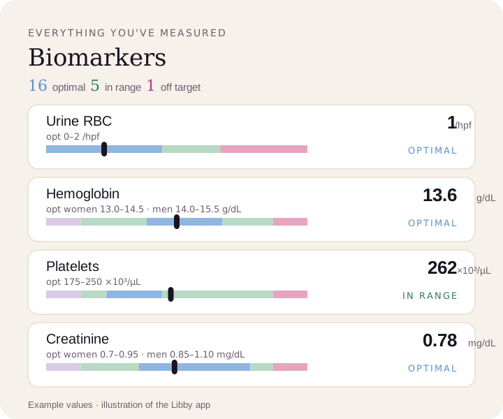
See your red-cell count next to the rest of your labs.
Upload a lab PDF or a photo of your report. Libby puts your urine red-cell count on your lab's range and the optimal range, next to your hemoglobin, platelets and creatinine, and tracks every new test.
View your urine RBCs ›