Blood in urine test: what a positive result means
A positive urine blood test can mean red cells, free hemoglobin or muscle pigment. What's normal, common causes, the neighbors to check, and when it's urgent.
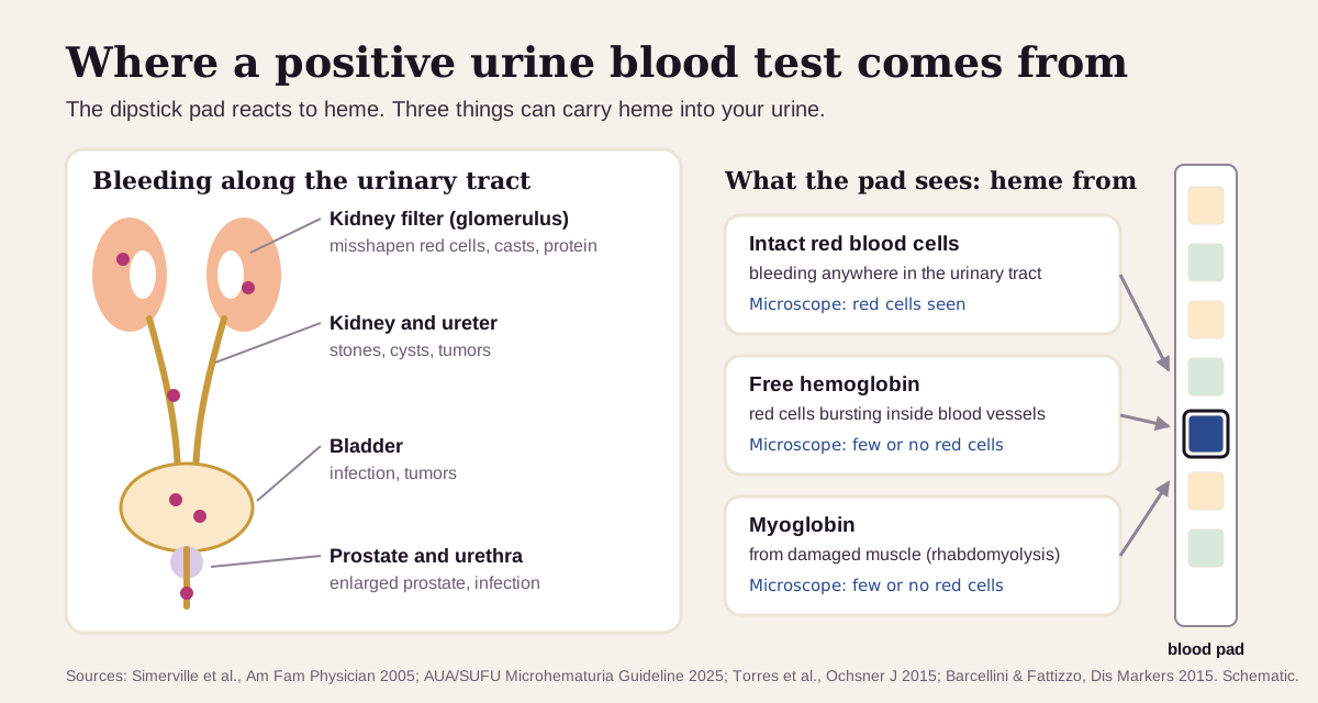
The urine blood test looks for heme, the iron-carrying part of hemoglobin, in your urine. A positive result usually means red blood cells are getting into your urine somewhere between your kidneys and the end of the urethra. Less often it means free hemoglobin from red cells that burst inside your blood vessels, or myoglobin from damaged muscle. A microscope count of red cells tells these apart.1
The short version
- Normal: a negative blood pad on the dipstick, and fewer than 3 red blood cells per high-power field (RBC/hpf) under the microscope. U.S. urology and internal-medicine guidelines call 3 or more "microhematuria".23
- A positive pad is a screen. It gets checked under the microscope. When the blood is confirmed, the causes found most often are urinary tract infection, an enlarged prostate and kidney stones, and in 43–68% of people no cause turns up.4 A hard workout, sex or a period can cause a temporary positive.5
- Visible blood (pink or red urine) always needs a clinician's look, even if it stops. Get care right away for clots, trouble passing urine, or fever with back or side pain.36
- Red cells or no red cells is the key split. A positive pad with red cells is bleeding (hematuria). A positive pad with few or no red cells points to free hemoglobin or muscle myoglobin.1
What the urine blood test measures
The "blood" pad on a urine dipstick reacts to heme. Heme acts like the enzyme peroxidase: it helps a chemical on the pad turn a dye blue, and more heme makes a darker pad.2 (More on the chemistry at the end.)
Three things carry heme into urine:1
- Red blood cells, from bleeding anywhere along the urinary tract. This is hematuria.
- Free hemoglobin, released when red cells burst inside your blood vessels (hemolysis). Once it outstrips haptoglobin, the blood protein that mops it up, the kidneys filter it into the urine.7
- Myoglobin, the oxygen-holding protein of muscle, released when muscle breaks down (rhabdomyolysis).8
The pad reads all three the same way. A microscope count of red cells separates them: red cells show up with hematuria and are few or absent with hemoglobin or myoglobin.1
Blood can be visible (gross hematuria: pink, red or reddish-brown urine) or microscopic (found only on testing). The blood test is part of a routine urinalysis, so many people first see it on a yearly checkup.5
What's a normal urine blood result?
Normal is a negative blood pad and 0–2 red cells per high-power field. The American Urological Association's guideline (AUA/SUFU, 2025) defines microhematuria as 3 or more red cells per high-power field on one properly collected sample, and the American College of Physicians uses the same line.23
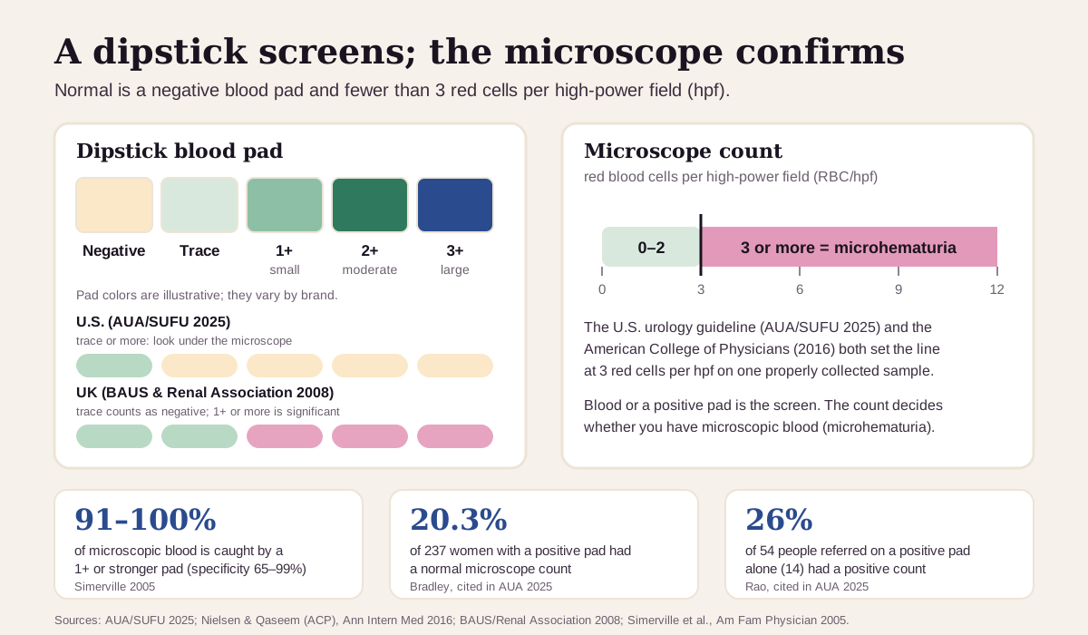
- The dipstick is sensitive but jumpy. A pad reading of 1+ or more catches 91–100% of microscopic blood, with a specificity of 65–99%.1 Up to 35% of positive pads turn out to be false alarms.4 In one series, 20.3% of 237 women with a positive pad had a normal microscope count. In another, only 14 of 54 people referred on a pad alone (26%) had a positive count.2
- That's why the U.S. rule has two steps. The AUA/SUFU says a pad reading of trace or more should prompt a microscope count, and only the count defines microhematuria.2 UK kidney and urology societies go the other way: they treat trace as negative, count 1+ or more as significant, and skip the microscope.9
- Microscopic blood is common. Screening studies of healthy volunteers found it in 2.4% to 31.1% of people, depending on who was tested.2
- How the sample is collected matters. The AUA/SUFU advises against testing the first morning urine or the first urine after vigorous exercise or sex, and says to retest after a period ends. Lots of flat squamous (skin) cells in the sample suggest contamination.2
Men and women share the same cut-offs. The AUA/SUFU uses sex and age only in its cancer-risk groups (below).2
"Optimal" urine blood: what health optimizers aim for
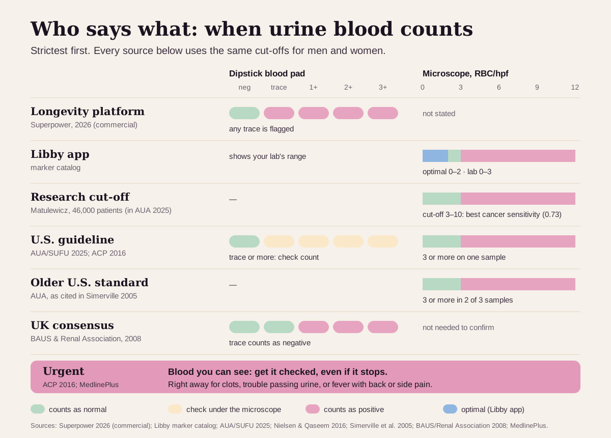
Everyone agrees on the target: no blood. For the dipstick there's no "better" within normal, and the Libby app shows your lab's range for it. Where sources differ is how small a signal counts as positive. Here's the ladder, strictest first:
| Target | Dipstick pad | Microscope count | Who uses it |
|---|---|---|---|
| Strictest (longevity platform) | negative; any trace flagged | not stated | Superpower's published optimal: "negative is optimal"10 |
| Libby app | your lab's range (negative) | optimal 0–2 RBC/hpf; lab range 0–3 | The optimal band shown in the Libby app for the red-cell count |
| Research cut-off | — | 3 or more | In over 46,000 patients, a cut-off of 3–10 RBC/hpf gave the best bladder-cancer sensitivity (0.73)2 |
| U.S. guideline | trace or more: get a microscope count | 3 or more on one sample | AUA/SUFU 2025 and the American College of Physicians23 |
| Older U.S. standard | — | 3 or more in 2 of 3 samples | The earlier AUA definition, as cited in 20051 |
| UK consensus | 1+ or more counts; trace is negative | not needed | British urology and kidney societies9 |
| Urgent | visible blood | visible blood | Any visible blood needs a clinician's look; clots, trouble passing urine, or fever with back or side pain need care right away36 |
Every source above uses the same cut-offs for men and women.
Why even a small amount gets checked:
- In a prospective study of more than 3,500 people referred for blood in their urine, urinary tract cancer turned up in 13.2% of those with visible blood and 3.1% of those with microscopic blood only.2
- In 1.2 million Israelis first examined at ages 16–25, persistent microscopic blood with no other finding was linked to 18.5 times the risk of kidney failure over about 22 years (34.0 vs 2.05 cases per 100,000 person-years). The absolute risk stayed low: 0.70% vs 0.045%.11
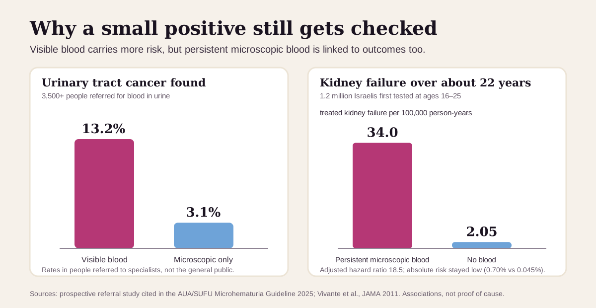
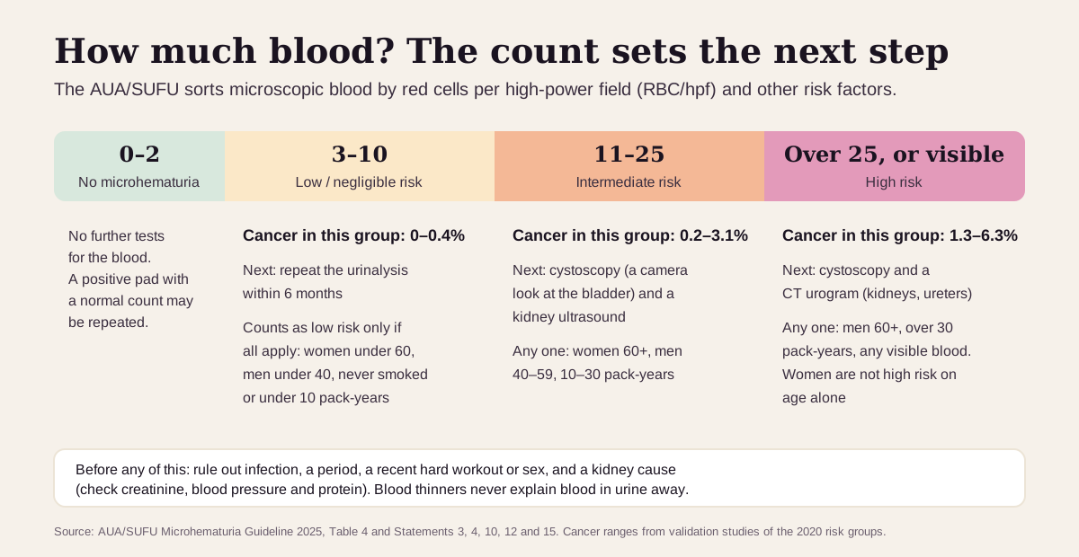
Keep "optimal" in perspective: a positive result is a reason to look, not a diagnosis. Most people with microscopic blood have nothing serious. In the AUA/SUFU's lowest-risk group, cancer turned up in 0–0.4%, so the guideline recommends a repeat urinalysis there rather than a camera test.2 The American College of Physicians also advises against urine tests to screen healthy adults for cancer.3 And there's no "too low" to worry about: negative is as good as this test gets.
What a positive urine blood test usually means
The first question is whether the blood is real and lasting. A positive pad is followed by a microscope count, and temporary causes are ruled out before anything else.24 Once microscopic blood is confirmed, the AUA/SUFU sorts people by the count and their risk factors:
Temporary causes: common, and they clear
- Hard exercise. Long-distance running is the classic example. A repeat urinalysis after 48–72 hours should be negative.1
- A period, sex, a viral illness, a minor injury or a recent catheter. Clinicians treat or wait out the cause and retest after at least 48 hours.45
- Contamination. Blood from the vagina, from bleeding hemorrhoids or in semen can end up in the sample.654
Confirmed microscopic blood: the usual suspects
In reviews of people with confirmed microscopic blood, the causes found were, roughly from most to least common:4
- No cause found: 43–68%.
- Urinary tract infection: 4–22%. Burning, fever, and a positive leukocyte-esterase or nitrite pad point to it. The blood should clear once the infection is treated, and guidelines ask for a repeat test to prove it.29
- Enlarged prostate (BPH): 10–13%.
- Kidney stones: 4–5%. Pain in the side (flank) can be a clue.2
- Bladder cancer: 2–4%. The risk is higher in men, after age 35, and in people who have smoked.4
- Less common: kidney cysts and kidney (glomerular) disease, about 2–3% each; kidney cancer, prostate cancer and a narrowed urethra, under 1% each.4
Blood thinners raise the chance of bleeding but never explain blood in urine away: guidelines ask for the same workup whether or not you take them.23
Visible blood
Visible blood carries the most weight. A 2005 review put the share of people with visible blood who have a urinary tract cancer at up to 20%,1 and the American College of Physicians advises urology referral for every adult with visible blood, even if it stopped on its own.3 Beets, rhubarb and some medicines can turn urine red without any blood.5
Positive pad, few or no red cells
- Myoglobin from muscle breakdown (rhabdomyolysis): tea-colored urine, with or without muscle pain. A CK blood test above about 5× the upper limit (around 1,000 IU/L) is the usual cut-off, and levels above 5,000 IU/L are closely tied to kidney damage.8
- Free hemoglobin from hemolysis: red cells bursting inside blood vessels. Low haptoglobin and high LDH (lactate dehydrogenase) on a blood test point to it.7
Get care now if you pass blood clots, are unable to urinate, or have fever, shaking chills, nausea or pain in your belly, side or back along with blood in your urine.6 Dark, tea-colored urine after extreme exertion or a crush injury, especially with muscle pain or weakness, also needs prompt care.8
Urine blood reads best next to its neighbors
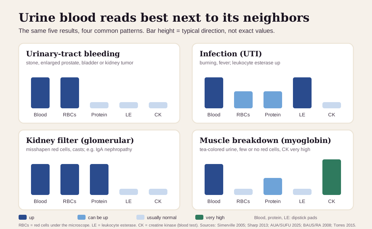
The pattern across your urinalysis and a couple of blood tests usually says more than the blood pad alone:17
| If you see… | It often points to… |
|---|---|
| Blood and red cells, protein about normal, no signs of infection | bleeding along the urinary tract: a stone, an enlarged prostate, or a bladder or kidney growth. Even heavy bleeding usually leaves the protein pad below 2+ |
| Blood and red cells with a positive leukocyte-esterase or nitrite pad, burning | urinary tract infection; retest after treatment |
| Blood, misshapen red cells or red-cell casts, and protein | the kidney's filter (glomerular disease); IgA nephropathy is the most common cause |
| Blood with few or no red cells, CK very high | myoglobin from muscle breakdown |
| Blood with few or no red cells, low haptoglobin, high LDH | free hemoglobin from hemolysis |
| Blood with many squamous cells, or during a period | contamination; repeat the sample |
When the kidney-filter pattern shows up, clinicians check creatinine, blood pressure and a urine protein or albumin ratio, and often bring in a kidney specialist alongside the urologist.294
How fast urine blood changes
Blood from a temporary cause clears fast. Exercise-related blood should be gone on a repeat test after 48–72 hours,1 and clinicians retest other temporary causes after at least 48 hours.4
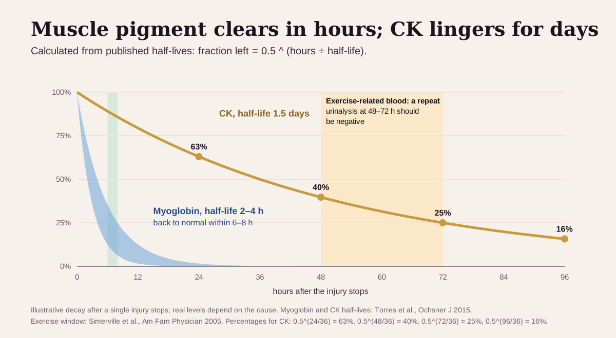
- Muscle pigment clears fast. Myoglobin in the blood is back to normal within 6–8 hours of a muscle injury, while CK stays up for days. A urine test a day later can miss it, which is why CK is the better test.8
- Hemoglobin clears fast too. After hemolysis, hemoglobin leaves the urine quickly, while iron deposits (hemosiderin) show up 3–4 days later and can last for weeks.7
- Bleeding comes and goes. A single urinalysis catches about 95% of people with microscopic blood. If a repeat after a temporary cause is negative, the AUA/SUFU says no further workup is needed for now. Blood that persists on repeat testing carries more weight: cancer was found in 0.35% vs 0.07% in a study of over 11,000 people.2
- Trends beat snapshots. A low-risk count gets a repeat urinalysis within 6 months. A count that rises, or new visible blood, restarts the workup.2
Going deeper: how the blood pad reads heme
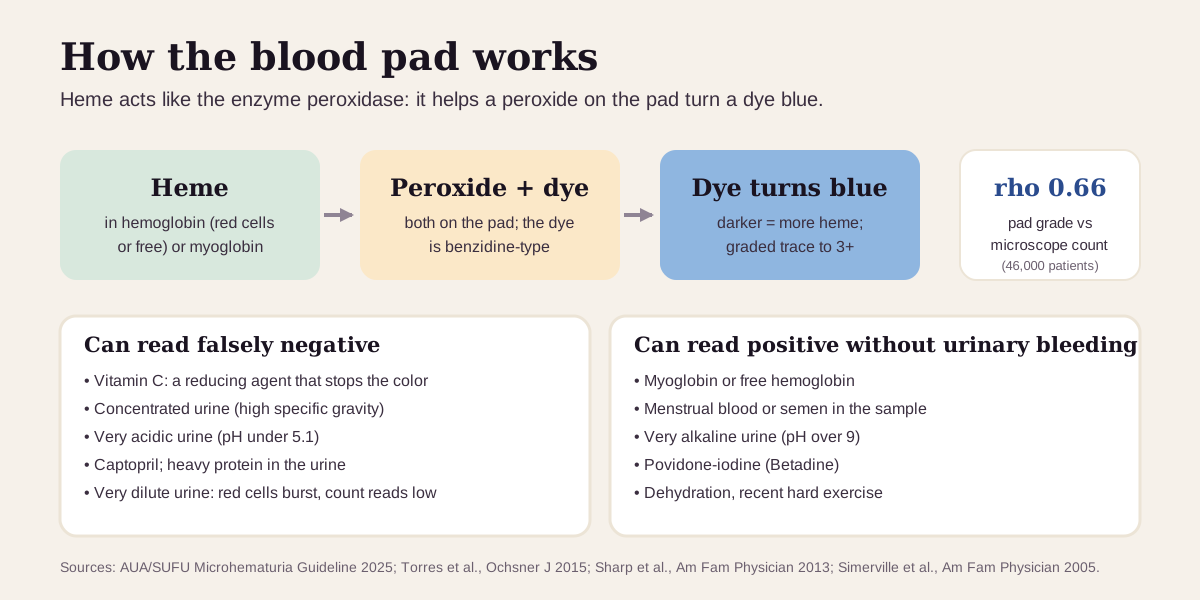
The pad holds a peroxide and a colorless benzidine-type dye. Heme speeds up the reaction between them, the way the enzyme peroxidase would, and the dye turns blue. Myoglobin does the same.28 More heme gives a darker color: in over 46,000 patients, the pad grade tracked the microscope count (rho 0.66).2
That chemistry explains the quirks:
- Vitamin C is a reducing agent. It interferes with the color and can make a bloody sample read negative, so people who take vitamin C supplements may do better with a microscope count first.4
- Concentration cuts both ways. Dehydration can push the pad positive, while a very high specific gravity, very acidic urine (pH under 5.1), heavy protein or the blood-pressure drug captopril can blunt it.1
- Things other than urinary-tract bleeding can turn the pad positive: myoglobin, free hemoglobin, menstrual blood, semen, very alkaline urine (pH over 9) and povidone-iodine.42
- Very dilute urine (below about 308 mOsm) bursts red cells before they're counted, so the microscope count can read low.2
Some reports split positives into "hemolyzed" and "non-hemolyzed". UK kidney and urology societies treat a 1+ of either kind as equally significant.9
Where the red cells came from. Red cells that squeeze through the kidney's filter often come out misshapen ("dysmorphic") and can clump into tube-shaped casts. With protein, that points to the kidney itself. Still, about 20% of people with biopsy-proven glomerulonephritis show blood alone.1
Questions worth asking
- Was the positive dipstick confirmed with a microscope count, and how many red cells per high-power field?
- Could exercise, sex, a period, an infection or a recent procedure explain it, and when should we repeat the test?
- Were red cells actually seen, or could this be hemoglobin or myoglobin?
- Do my protein, creatinine and blood pressure point toward the kidney?
- Given my age, sex and smoking history, which AUA risk group am I in, and what does that mean for next steps?
- I saw blood once and it went away. Should I still see a urologist?
FAQ
What does trace blood in urine mean? A trace reading is the faintest positive. U.S. guidelines ask for a microscope count to confirm it; UK guidelines treat trace as negative.29 Many positive pads turn out to be temporary or false alarms.4
Can exercise cause blood in urine? Yes. Hard exercise, especially long-distance running, is a common, harmless cause. A repeat test after 48–72 hours should be negative.1
Is blood in urine a sign of cancer? Usually it has another cause. In people referred for it, cancer turned up in about 3% with microscopic blood only and about 13% with visible blood. The risk rises with age, male sex and smoking.24
Why is my dipstick positive when no red cells were seen? The pad reacts to heme in free hemoglobin and muscle myoglobin as well as red cells. A CK test checks for muscle breakdown, and haptoglobin and LDH check for hemolysis. Very dilute urine can also burst red cells before they're counted.1872
This guide is for education. It can't diagnose you or replace care from a clinician who knows your history.
Footnotes
-
Simerville JA, Maxted WC, Pahira JJ. Urinalysis: a comprehensive review. Am Fam Physician. 2005;71(6):1153–1162. Dipstick chemistry and accuracy, false positives and negatives, the older AUA definition, and the glomerular vs urologic patterns. AAFP ↩ ↩2 ↩3 ↩4 ↩5 ↩6 ↩7 ↩8 ↩9 ↩10 ↩11 ↩12 ↩13 ↩14 ↩15 ↩16 ↩17 ↩18 ↩19
-
Barocas DA, Lotan Y, Matulewicz RS, et al. Updates to Microhematuria: AUA/SUFU Guideline (2025). J Urol. 2025;213(5):547–557. The U.S. urology guideline (first published 2020, amended 2025). It defines microhematuria as 3 or more RBC/hpf, sets the risk groups (Table 4), and summarizes the dipstick, collection and outcome studies quoted here. AUA · PubMed ↩ ↩2 ↩3 ↩4 ↩5 ↩6 ↩7 ↩8 ↩9 ↩10 ↩11 ↩12 ↩13 ↩14 ↩15 ↩16 ↩17 ↩18 ↩19 ↩20 ↩21 ↩22 ↩23 ↩24 ↩25 ↩26 ↩27 ↩28 ↩29 ↩30 ↩31 ↩32
-
Nielsen M, Qaseem A; High Value Care Task Force of the American College of Physicians. Hematuria as a Marker of Occult Urinary Tract Cancer: Advice for High-Value Care From the American College of Physicians. Ann Intern Med. 2016;164(7):488–497. Confirm a positive pad with 3 or more RBC/hpf; refer every adult with visible blood. PubMed ↩ ↩2 ↩3 ↩4 ↩5 ↩6 ↩7 ↩8 ↩9 ↩10
-
Sharp VJ, Barnes KT, Erickson BA. Assessment of asymptomatic microscopic hematuria in adults. Am Fam Physician. 2013;88(11):747–754. How often each cause is found, and dipstick pitfalls including vitamin C. AAFP ↩ ↩2 ↩3 ↩4 ↩5 ↩6 ↩7 ↩8 ↩9 ↩10 ↩11 ↩12 ↩13 ↩14 ↩15 ↩16
-
National Library of Medicine. Blood in Urine. MedlinePlus. Plain-language causes, foods that color urine, and collection tips. MedlinePlus ↩ ↩2 ↩3 ↩4 ↩5
-
National Library of Medicine. Urine - bloody. MedlinePlus Medical Encyclopedia. Reviewed July 2025. When to contact a clinician right away. MedlinePlus ↩ ↩2 ↩3 ↩4 ↩5
-
Barcellini W, Fattizzo B. Clinical Applications of Hemolytic Markers in the Differential Diagnosis and Management of Hemolytic Anemia. Dis Markers. 2015;2015:635670. Haptoglobin, LDH, and how free hemoglobin reaches the urine. PMC ↩ ↩2 ↩3 ↩4 ↩5 ↩6
-
Torres PA, Helmstetter JA, Kaye AM, Kaye AD. Rhabdomyolysis: pathogenesis, diagnosis, and treatment. Ochsner J. 2015;15(1):58–69. CK cut-offs, myoglobin and CK half-lives, and why myoglobin turns the blood pad positive. PMC ↩ ↩2 ↩3 ↩4 ↩5 ↩6 ↩7 ↩8 ↩9
-
Anderson J, Fawcett D, Feehally J, et al. Joint Consensus Statement on the Initial Assessment of Haematuria. Renal Association and British Association of Urological Surgeons; July 2008. The UK definitions: trace counts as negative, 1+ or more is significant, and microscopy is not needed to confirm. BAUS (PDF) ↩ ↩2 ↩3 ↩4 ↩5 ↩6 ↩7 ↩8 ↩9
-
Superpower. Occult Blood (Urine) Test. Updated September 1, 2026. States "The optimal result is negative; any presence warrants investigation." A commercial source, listed as what the optimizer community uses. superpower.com ↩ ↩2
-
Vivante A, Afek A, Frenkel-Nir Y, et al. Persistent asymptomatic isolated microscopic hematuria in Israeli adolescents and young adults and risk for end-stage renal disease. JAMA. 2011;306(7):729–736. 1,203,626 people aged 16–25, followed about 22 years. PubMed ↩ ↩2
Educational content, not medical advice. Libby is a personal record tool, not a medical service — it doesn't diagnose, treat, or prescribe. Reference ranges vary by lab and by person. Talk to a qualified healthcare professional about your results.
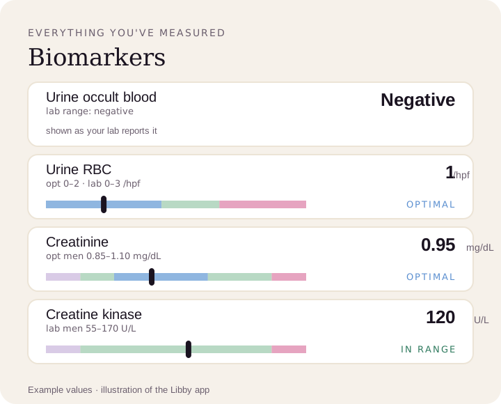
See every result against your lab's range.
Upload a lab PDF or a photo of your report. Libby shows your urine blood result as your lab reports it, next to your red-cell count, creatinine and more, and tracks each new test.
View your urine blood ›